
Psychoda laticaula Quate
|
publication ID |
https://doi.org/10.5281/zenodo.205242 |
|
publication LSID |
lsid:zoobank.org:pub:EC72D183-3ED9-4DF3-AE7D-32C469575224 |
|
persistent identifier |
https://treatment.plazi.org/id/03A8E828-FF92-2905-DCB5-77C5FD8EFD9C |
|
treatment provided by |
Plazi (2016-04-11 21:29:21, last updated 2019-10-18 19:40:02) |
|
scientific name |
Psychoda laticaula Quate |
| status |
|
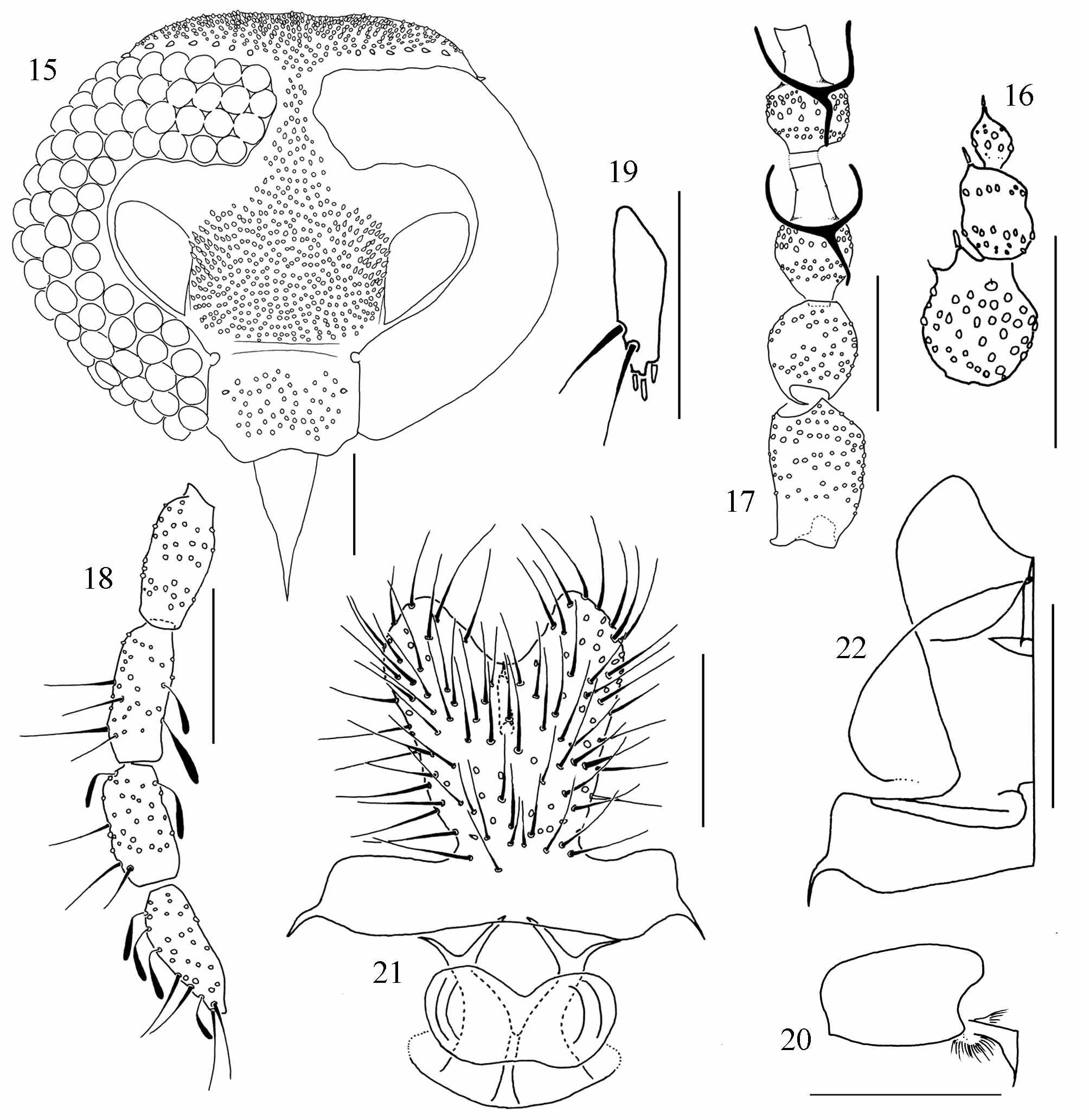
( Figs 15–22 View FIGURES 15 – 22 )
Psychoda laticaula Quate, 1996: 67 . Type locality: Costa Rica (Limón, Puerto Viejo de Talamanca )
Diagnosis. Antenna with 14 flagellomeres, 12 th and 13 th fused, 14 th separated and shorter; palpal formula 1.0: 0.9: 0.9:1.0; subgenital plate heart shaped, with basal band and a large semi-circular structure internally.
Female. Head ( Fig. 15 View FIGURES 15 – 22 ): vertex, frons and clypeus pilose; hair patch of frons extending to facet row 1 or meeting the hair patch of vertex; eye bridge with 4 facets; eyes separated by 1.0– 1.5 facet diameters; 6-7 supra-ocular setae; interocular suture absent; clypeus with 1 stronger lateral scar; frontoclypeal suture absent, sometimes with a weak band linking the tentorial fossets; antenna with 14 flagellomeres, the 3 apical reduced ( Fig. 16 View FIGURES 15 – 22 ); 12 th and 13 th flagellomeres fused, 14 th shorter and separated from 13 th; spines presents on 11 th, 13 th and 14 th flagellomeres; scape cylindrical, 1.5 the length of the subspherical pedicel ( Fig. 17 View FIGURES 15 – 22 ); ascoids in Y ( Fig. 17 View FIGURES 15 – 22 ); palpal formula 1.0: 0.9: 0.9:1.0 ( Fig. 18 View FIGURES 15 – 22 ); labellum with 3 apical teeth, 1 subapical tooth and 2 lateral setae ( Fig. 19 View FIGURES 15 – 22 ). Wing: Sc vein not extending beyond base of vein Rs; radial and medial forks complete. Distitarsus with apical projection ( Fig. 20 View FIGURES 15 – 22 ). Terminalia: subgenital plate homogeneously pilose, longer than wide, bilobed, heart shaped, with well developed naked basal band ( Fig. 21 View FIGURES 15 – 22 ); genital digit present; subgenital plate internally with a lateral line curved to the apical concavity and a semicircular, slightly sclerotized structure ( Fig. 22 View FIGURES 15 – 22 ); genital chamber short, as illustrated ( Fig. 21 View FIGURES 15 – 22 ).
Male. Unknown.
Material examined. Type material: holotype Ƥ COSTA RICA, Limón, Puerto Viejo de Talamanca , Sealevel, 20-22.vii. 1993, Light trap, col. L. W. Quate ( INBio); paratype Ƥ COSTA RICA, Limón, Puerto Viejo de Talamanca , Sealevel, 20.vii. 1993, Light trap, col. L. W. Quate ( USNM). Other specimens: 1 Ƥ BRAZIL, Bahia, Ituberá, Reserva Michelin, Pancagê, 08–09.vi. 2007, Luminosa, col. E. Alvim & J. Oliveira (MZUEFS).
Distribution. Costa Rica, Nicaragua ( Collantes & Martinez-Ortega 1999), Brazil (Bahia).
Comments. The wings of the Brazilian specimen were damaged during preparation, so they were not measured. The right palpus of the holotype has only 3 segments, the third and forth segments are apparently fused. The morphology of antenna apex and ascoids on this species is close to what is found in the subgenus Psychomora , but the 11 th and 12 th flagellomeres are not fused as in the description of Psychomora provided by Ježek (1984).This is the first record of this species in Brazil.
Collantes, F. & Martinez-Ortega, E. (1999) Nuevas citas de especies conocidas de Psychodinae (Diptera, Psychodidae) em Nicaragua. Revista Nicaraguense de Entomologia, 48, 17 - 27.
Jezek, J. (1984) Six new genera of the tribe Psychodini End. (Diptera, Psychodidae). Acta Faunistica Entomologica Musei Nationalis Pragae, 17, 133 - 153.
Quate, L. W. (1996) Preliminary taxonomic of Costa Rica: Psychodidae (Diptera), exclusive of Plebotominae. Revista de Biologia Tropical, 44 (Supplement 1), 1 - 81
No known copyright restrictions apply. See Agosti, D., Egloff, W., 2009. Taxonomic information exchange and copyright: the Plazi approach. BMC Research Notes 2009, 2:53 for further explanation.
|
Kingdom |
|
|
Phylum |
|
|
Class |
|
|
Order |
|
|
Family |
|
|
Genus |
Psychoda laticaula Quate
| Cordeiro, Danilo, Bravo, Freddy & De, Claudio J. B. 2011 |
Psychoda laticaula
| Quate 1996: 67 |
1 (by plazi, 2016-04-11 21:29:21)
2 (by ImsDioSync, 2016-12-15 21:56:10)
3 (by ImsDioSync, 2016-12-15 21:59:30)
4 (by ImsDioSync, 2017-06-17 03:32:33)
5 (by ImsDioSync, 2017-06-17 04:11:06)
6 (by ExternalLinkService, 2019-09-26 20:05:45)
7 (by ExternalLinkService, 2019-10-09 06:47:58)
8 (by ExternalLinkService, 2019-10-09 06:53:15)
9 (by ExternalLinkService, 2019-10-18 09:42:51)
10 (by ExternalLinkService, 2019-10-18 19:40:02)
11 (by ExternalLinkService, 2019-10-18 23:29:10)
12 (by ExternalLinkService, 2019-10-19 18:46:58)
13 (by ExternalLinkService, 2019-10-19 20:35:48)
14 (by ExternalLinkService, 2022-01-30 15:00:57)
15 (by ExternalLinkService, 2022-02-20 21:02:41)
16 (by plazi, 2023-10-26 01:14:04)