
Psychoda laticaula Quate
|
publication ID |
https://doi.org/10.5281/zenodo.205242 |
|
publication LSID |
lsid:zoobank.org:pub:EC72D183-3ED9-4DF3-AE7D-32C469575224 |
|
persistent identifier |
https://treatment.plazi.org/id/03A8E828-FF92-2905-DCB5-77C5FD8EFD9C |
|
treatment provided by |
Plazi |
|
scientific name |
Psychoda laticaula Quate |
| status |
|
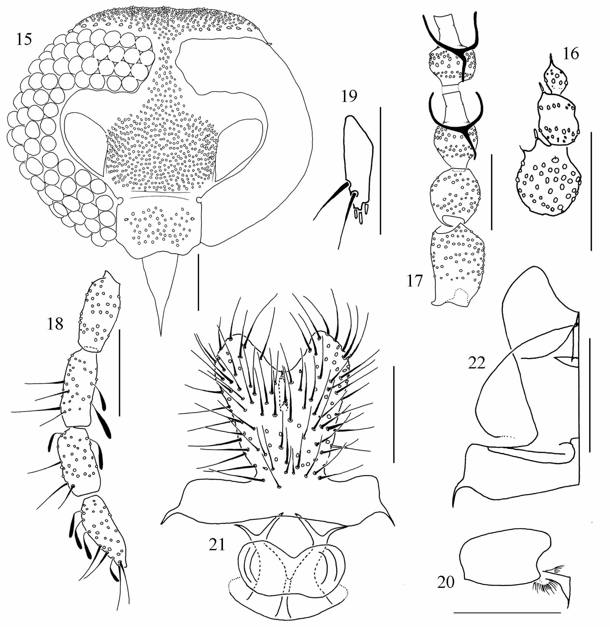
( Figs 15–22 View FIGURES 15 – 22 )
Psychoda laticaula Quate, 1996: 67 . Type locality: Costa Rica (Limón, Puerto Viejo de Talamanca )
Diagnosis. Antenna with 14 flagellomeres, 12 th and 13 th fused, 14 th separated and shorter; palpal formula 1.0: 0.9: 0.9:1.0; subgenital plate heart shaped, with basal band and a large semi-circular structure internally.
Female. Head ( Fig. 15 View FIGURES 15 – 22 ): vertex, frons and clypeus pilose; hair patch of frons extending to facet row 1 or meeting the hair patch of vertex; eye bridge with 4 facets; eyes separated by 1.0– 1.5 facet diameters; 6-7 supra-ocular setae; interocular suture absent; clypeus with 1 stronger lateral scar; frontoclypeal suture absent, sometimes with a weak band linking the tentorial fossets; antenna with 14 flagellomeres, the 3 apical reduced ( Fig. 16 View FIGURES 15 – 22 ); 12 th and 13 th flagellomeres fused, 14 th shorter and separated from 13 th; spines presents on 11 th, 13 th and 14 th flagellomeres; scape cylindrical, 1.5 the length of the subspherical pedicel ( Fig. 17 View FIGURES 15 – 22 ); ascoids in Y ( Fig. 17 View FIGURES 15 – 22 ); palpal formula 1.0: 0.9: 0.9:1.0 ( Fig. 18 View FIGURES 15 – 22 ); labellum with 3 apical teeth, 1 subapical tooth and 2 lateral setae ( Fig. 19 View FIGURES 15 – 22 ). Wing: Sc vein not extending beyond base of vein Rs; radial and medial forks complete. Distitarsus with apical projection ( Fig. 20 View FIGURES 15 – 22 ). Terminalia: subgenital plate homogeneously pilose, longer than wide, bilobed, heart shaped, with well developed naked basal band ( Fig. 21 View FIGURES 15 – 22 ); genital digit present; subgenital plate internally with a lateral line curved to the apical concavity and a semicircular, slightly sclerotized structure ( Fig. 22 View FIGURES 15 – 22 ); genital chamber short, as illustrated ( Fig. 21 View FIGURES 15 – 22 ).
Male. Unknown.
Material examined. Type material: holotype Ƥ COSTA RICA, Limón, Puerto Viejo de Talamanca , Sealevel, 20-22.vii. 1993, Light trap, col. L. W. Quate ( INBio); paratype Ƥ COSTA RICA, Limón, Puerto Viejo de Talamanca , Sealevel, 20.vii. 1993, Light trap, col. L. W. Quate ( USNM). Other specimens: 1 Ƥ BRAZIL, Bahia, Ituberá, Reserva Michelin, Pancagê, 08–09.vi. 2007, Luminosa, col. E. Alvim & J. Oliveira (MZUEFS).
Distribution. Costa Rica, Nicaragua ( Collantes & Martinez-Ortega 1999), Brazil (Bahia).
Comments. The wings of the Brazilian specimen were damaged during preparation, so they were not measured. The right palpus of the holotype has only 3 segments, the third and forth segments are apparently fused. The morphology of antenna apex and ascoids on this species is close to what is found in the subgenus Psychomora , but the 11 th and 12 th flagellomeres are not fused as in the description of Psychomora provided by Ježek ( 1984).This is the first record of this species in Brazil.
No known copyright restrictions apply. See Agosti, D., Egloff, W., 2009. Taxonomic information exchange and copyright: the Plazi approach. BMC Research Notes 2009, 2:53 for further explanation.
|
Kingdom |
|
|
Phylum |
|
|
Class |
|
|
Order |
|
|
Family |
|
|
Genus |
Psychoda laticaula Quate
| Cordeiro, Danilo, Bravo, Freddy & De, Claudio J. B. 2011 |
Psychoda laticaula
| Quate 1996: 67 |