
Saccogaster parva Cohen & Nielsen, 1972
|
publication ID |
https://doi.org/ 10.5281/zenodo.208677 |
|
publication LSID |
lsid:zoobank.org:pub:7CC3476C-AC83-4401-8236-53F59CB88C8F |
|
DOI |
https://doi.org/10.5281/zenodo.6175256 |
|
persistent identifier |
https://treatment.plazi.org/id/EB64193E-E22A-A359-86A8-FEEEFD5F0F1E |
|
treatment provided by |
Plazi |
|
scientific name |
Saccogaster parva Cohen & Nielsen, 1972 |
| status |
|
Saccogaster parva Cohen & Nielsen, 1972 View in CoL
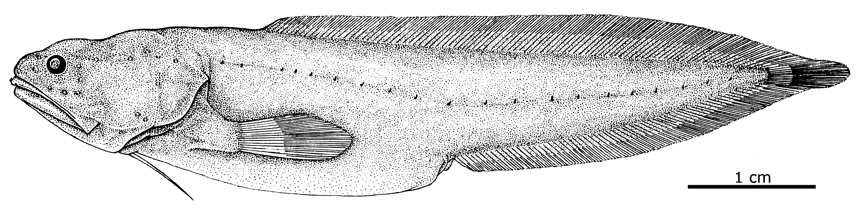
Figs 13 View FIGURE 13 , 14 View FIGURE 14 , 26 View FIGURE 26 , Table 1 View TABLE 1 , 2, 5 View TABLE 5
Saccogaster parva Cohen & Nielsen 1972: 459 View in CoL , fig. 5 (type locality: off southern Brazil). Saccogaster parva: Nielsen et al. 1999: 110 View in CoL .
Material examined (1 specimen, 58 mm SL). Holotype: ZMH 25268, female, 58 mm SL, off southern Brazil, 24°21’S, 43°54’W, R/V Walther Herwig, st. 90/68, bottom trawl, 500 m, 2 Mar. 1968.
Diagnosis. Saccogaster parva differs from all other Saccogaster species in having 5–6 developed rakers on anterior gill arch and no spines on frontal plate and ethmoid. Also the following combination of characters is diagnostic: Skin thin and transparent; numerous neuromasts below translucent head skin arranged in five distinct clusters; scales absent; three developed rakers on anterior gill arch 3–4 times length of gill filaments; palatines with one row of teeth; antero-ventrally directed spine on lower angle of preoperculum; prolonged pectoral peduncle; precaudal vertebrae 16 and total vertebrae 54; fin rays in dorsal 91, anal 64 and pectoral 14.
Similarity. Saccogaster parva and S. nikoliviae are the only species with numerous, developed neuromasts on the head, but they differ in other characters such as number of developed gill rakers 6 vs. 3 in S. nikoliviae , absence of scales vs. present in S. nikoliviae and no spines behind eyes or on ethmoid vs. present in S. nikoliviae .
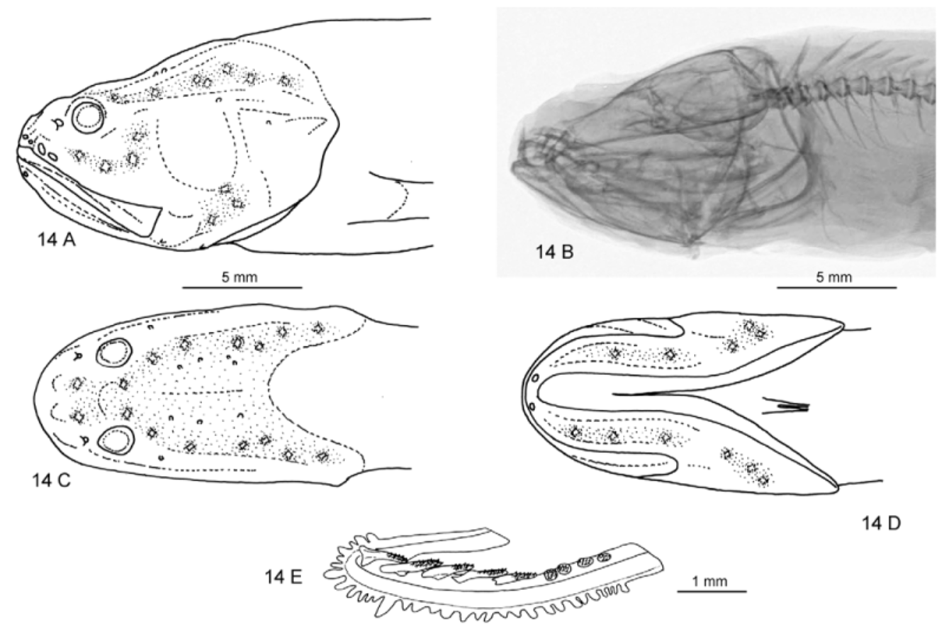
Description. The principal meristic and morphometric characters are shown in Table 5 View TABLE 5 . Body compressed and relatively short. Scales absent. Lateral line continuous, originating above preopercle descending gradually to midline over anus and ending close to basis of caudal fin; ca. 20 lateral line papillae. Dorsal fin origin above proximal part of pectoral fin and anal fin origin well behind midpoint of fish. Pectoral fins end halfway to anus. Pectoral peduncle prolonged. Head compressed with blunt snout. Opercular spine flat, preopercular spine hidden. Posteror, vertically expanded part of maxillary strongly sheathed dorsally. Anterior gill arch ( Fig. 14 View FIGURE 14 E) with two knobs on upper branch, a developed raker in angle, and lower branch with 5–6 developed rakers followed by 6–8 knobs. Pseudobranchial filaments 2. Ovaries with developing eggs.
Axial skeleton (from radiographs): Number of precaudal vertebrae 16. Anterior neural spine 1/3 length of second spine. Neural spines 2–5 decreasing in length and with pointed tips. Neural spines 6–10 with blunt tips and all remaining neural and haemal spines with pointed tips. Parapophyses developed on vertebrae 7–16. Pleural ribs on vertebrae 2–11 and epipleural ribs seen on vertebrae 3–6.
Dentition: Vomer boomerang-shaped with about six fangs and a scattering of smaller teeth. Palatines with one row of fang-like teeth. Dentaries with an irregular row of about 15 fangs and an outer narrow band of shorter teeth. Premaxillaries with a narrow band of small, granular teeth and five needle-like teeth at the symphysis
Head morphology ( Fig. 14 View FIGURE 14 A–D): Head profile straight above eyes. Head without spines on frontal plate and on ethmoid, but blunt, sub-dermal spine above eyes. Fig. 14 View FIGURE 14 B shows a conspicuous thickening of the frontal at the location where spines would be expected, indicating that larger specimens might in fact have small frontal spines. Also a broad hump is indicated on the snout at the position where a (cartilaginous) spine might be expected. Anterior nostril placed close to upper lip; posterior nostril moderately large with anterior flap, placed close to eye. Head pores: 2 supraorbital pores near tip of snout, 1 anterior infraorbital pore below and in front of eye, 1 anterior mandibular pore at tip of jaw. Head skin thin, tight. Total of 17–18 diamond-shaped, light-colored neuromasts visible below transparent skin arranged in five clusters: 4 infraorbital along upper lip and curving around eye, 2 pairs between eyes, one of them slightly in front of eyes, 6 supraorbital along line above opercle and behind eyes, 2–3 mandibular along jaw, 3 on the lower preopercle. Head with dusky background pigmentation chiefly around neuromasts. Upper jaw ends well behind eye; posterior end of maxilla vertically expanded. Opercular spine pointed but flat, subdermal, not reaching hind margin of opercle. Small, subdermal anterior-ventrally pointed spine at lower angle of preoperculum.
Otolith: Dissolved.
Coloration: Violet when caught.
Biology and distribution ( Fig. 26 View FIGURE 26 ). Caught on the bottom on the upper continental slope at a depth of 500 m. Known only from the holotype off southern Brazil.

| ZMH |
Zoologisches Museum Hamburg |
No known copyright restrictions apply. See Agosti, D., Egloff, W., 2009. Taxonomic information exchange and copyright: the Plazi approach. BMC Research Notes 2009, 2:53 for further explanation.
|
Kingdom |
|
|
Phylum |
|
|
Class |
|
|
Order |
|
|
Family |
|
|
Genus |
Saccogaster parva Cohen & Nielsen, 1972
| Nielsen, Jørgen G., Schwarzhans, Werner & Cohen, Daniel M. 2012 |
Saccogaster parva
| Nielsen 1999: 110 |
| Cohen 1972: 459 |