
Pardosa tuberosa, Wang, Dong & Zhang, Zhi-Sheng, 2014
|
publication ID |
https://doi.org/10.11646/zootaxa.3856.2.4 |
|
publication LSID |
lsid:zoobank.org:pub:5C95C6B3-DD2A-4D10-A698-9761F5AA96A1 |
|
DOI |
https://doi.org/10.5281/zenodo.6136168 |
|
persistent identifier |
https://treatment.plazi.org/id/193F878A-7627-FFF7-D8BF-FD04FD5B6511 |
|
treatment provided by |
Plazi (2016-04-17 10:49:14, last updated 2024-11-28 04:02:02) |
|
scientific name |
Pardosa tuberosa |
| status |
sp. nov. |
Pardosa tuberosa View in CoL sp. nov
Figs 3 View FIGURE 3 A–E, 4A–H; 10
Type material. Holotype Male, China, Yunnan Province: Xishuangbanna Dai Autonomous Prefecture, Jinghong City, Mengla County, near Mannasan Bridge, 21°26.26′ N, 101°33.47′ E, alt. 632 m, 26 May 2011, Z.X. Li & G.C. Zhou leg. ( SWUC). Paratypes: 1 male and 2 females, with same data as holotype ( SWUC); 4 males and 5 females, Pu’er Prefecture, Simao District, Manxie tunnel, 22°43.85′ N, 100°56.24′ E, alt. 1201 m, 29 May 2011, Z.X. Li & G.C. Zhou leg. ( SWUC); 1 male, Puer City, Simao District, Meizihu Park, 22°45.25′ N, 100°59.04′ E, alt. 1339 m, 21 May 2011, Z.X. Li & G.C. Zhou leg. ( SWUC); 1 male and 1 female, Ruili City, Nansang Village, rubber plantation, 24°1.47′ N, 97°49.14′ E, alt. 820 m, 4 June 2011, Z.X. Li & G.C. Zhou leg. ( SWUC); 1 female, Xishuangbanna Dai Autonomous Prefecture, Jinghong City, Mengla County, Mohan, 21°11.18′ N, 101°41.12′ E, alt. 889 m, 27 May 2011, Z.X. Li & G.C. Zhou leg. ( SWUC).
Etymology. The specific name comes from the Latin words ‘ tuberosus ’, meaning ‘full of protuberances’ and refers to the top of distal haematodocha with lots of protuberances; adjective.
Diagnosis. The new species can be distinguished from all other Pardosa nebulosa -group species by the presence of the many protuberances on the surface of distal haematodocha in the ventral view of male palp. The new species is also similar to P. chapini ( Fox, 1935) ( Figs 8 View FIGURE 8 A–H, 9A–D, widely distrubuted in South China) in having the similar shape of palea, a long stem of septum and large spermathecae, but differs from the latter by the longer than wide median apophysis, the slightly curved anterior margin of tegulum, the narrow distance of hoods, the slender stem of septum and the stronger spermathecae. It is also similar to P. pseudochapini Peng, 2011 (= P. bidentata in Qu et al., 2010: 388, figs 1a–b, 7a–d) (found in the same area) in having a similarly shaped median apophysis, the same position of subtegulum, a long stem of septum and large spermathecae, but can be distinguished by the irregular shape of palea, the slightly protruded anterior margin of tegulum, the absence of "two crenated divarications", the slender stem of septum and stronger spermathecae.
Description. Male total length 5.50–6.46. Holotype ( Figs 4 View FIGURE 4 A, 4C) total length 6.46. Prosoma 3.26 long, 2.67 wide; opisthosoma 2.93 long, 1.60 wide. Carapace yellowish brown, with light brown submarginal bands. Median band brownish-yellow with branched sides, dilated in cephalic area. Lateral bands dark brown and broad. Eye region black. Flanks of the head region steep in frontal view. Fovea vertical. Cervical groove and radial furrows indistinct. Eye sizes and interdistances: AME 0.49, ALE 0.46, PME 1.55, PLE 1.23; AME–AME 0.19, AME–ALE 0.23, PME–PME 1.21, PME–PLE 1.06. Clypeus height 0.89. Chelicerae elongate, black brown, with two promarginal and three retromarginal teeth. Labium and endites grey, longer than wide. Sternum grey, heart-shaped, with sparse black hairs. Legs yellow. Leg measurements: I 7.22 (1.73, 2.38, 2.00, 1.11); II 5.64 (1.63, 1.84, 1.50, 0.67); III 5.47 (1.55, 1.64, 1.53, 0.75); IV 7.89 (2.03, 2.35, 2.75, 1.16). Leg formula: 4123. Opisthosoma long oval. Dorsum dark brown, with distinct yellow cardiac mark in anterior half part, and with black irregular markings in posterior half part. Venter of opisthosoma yellow, with small, yellow spinnerets.
Male palp ( Figs 3 View FIGURE 3 A–C, 4D–H) with a large tegulum. Subtegulum rounded. Median apophysis with an arm, horizontally extending retrolaterally, strongly curved distally. Many protuberances located on the surface of distal haematodocha. Embolus slender, originating on the prolatero-basal side of palea. Terminal apophysis hook-like.
Female paratype ( Fig. 4 View FIGURE 4 B) total length 6.82. Prosoma 3.16 long, 2.62 wide; opisthosoma 3.43 long, 2.14 wide. Eye sizes and interdistances: AME 0.36, ALE 0.31, PME 1.03, PLE 1.10; AME–AME 0.27, AME–ALE 0.20, PME–PME 0.97, PME–PLE 1.43. Clypeus height 0.84. Leg measurements: I 8.43 (2.32, 2.93, 2.05, 1.13); II 5.76 (1.73, 1.87, 1.43, 0.73); III 5.29 (1.51, 1.70, 1.40, 0.68); IV 12.10 (3.04, 3.63, 3.82, 1.61). Leg formula: 4123. Colouration and pattern similar to that of male, but opisthosoma relatively darker than that of male.
Epigyne ( Figs 3 View FIGURE 3 D–E, 4I –J) with a pair of hoods anteriorly, which closely near each other. Anchor-like septum with a slender stem and a much wider than long base. Spermathecae scleritised, with a rounded base and a thin head. Fertilisation ducts transparent, posteriorly originating from the inner side of spermathecae.
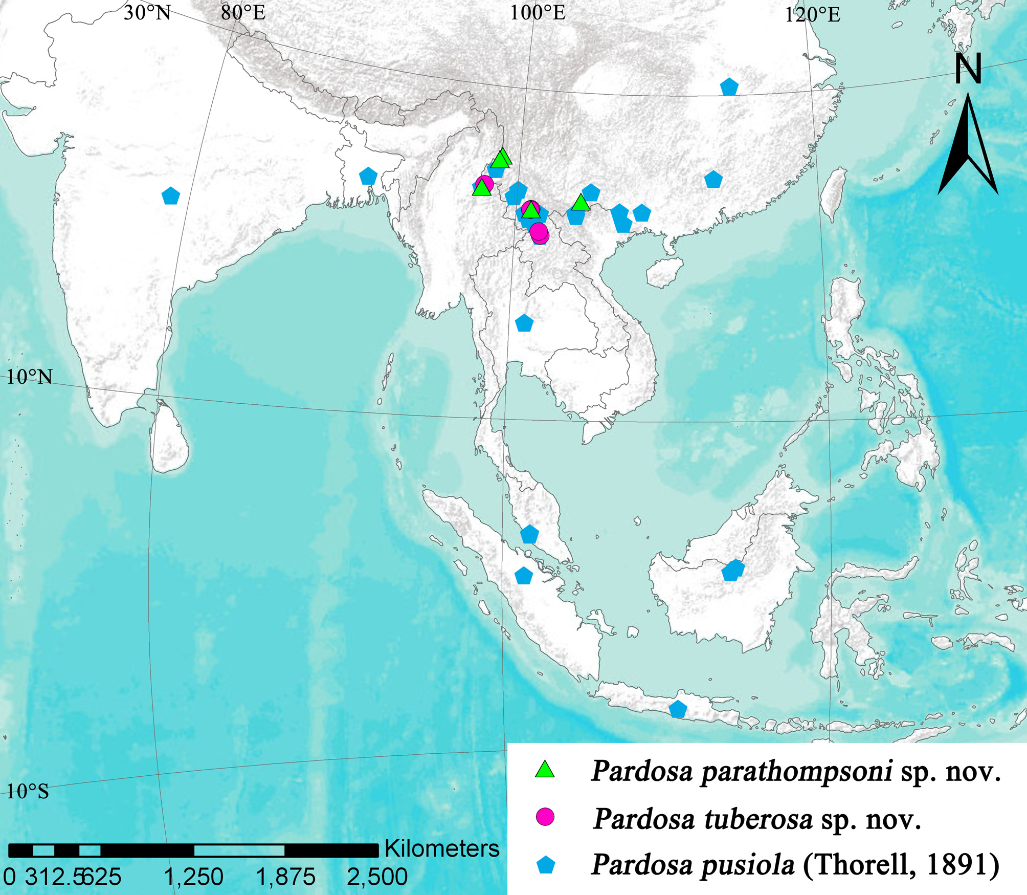
Distribution. China (Yunnan) ( Fig. 10 View FIGURE 10 )
Fox, I. (1935) Chinese spiders of the family Lycosidae. Journal of Washington Academy of Science 25, 451 - 456.
Peng, X. J. (2011) A new name of Pardosa bidentata Qu, Peng & Yin, 2010 (Araneae: Lycosidae). Acta arachnologica Sinica, 20, 9.
Qu, L. L., Peng X. J. & Yin, C. M. (2010) Six new species of the spider genus Pardosa (Araneae: Lycosidae) from Yunnan, China. Oriental Insects, 44, 387 - 404. http: // dx. doi. org / 10.1080 / 00305316.2010.10417623
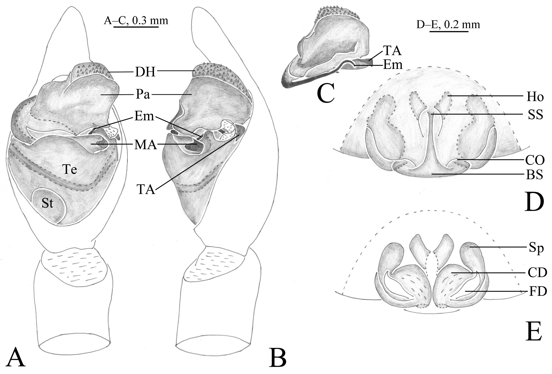
FIGURE 3. Pardosa tuberosa sp. nov., Male holotype (A, B) and female paratype (D, E) from the area near Mannasan Bridge, male paratype (C) from Manxie tunnel. A. Left male palp, ventral view; B. Same, retrolateral view; C. Apical part of bulb, ventral view; D. Epigyne, ventral view; E. Vulva, dorsal view. Abbreviations: BS, base of septum; CD, copulatory duct; CO, copulatory opening; DH, distal haematodocha; Em, embolus; FD, fertilization duct; Ho, hood; MA, median apophysis; Pa, palea; Sp, spermathecae; SS, stem of septum; St, subtegulum; TA, Terminal apophysis; Te, tegulum.
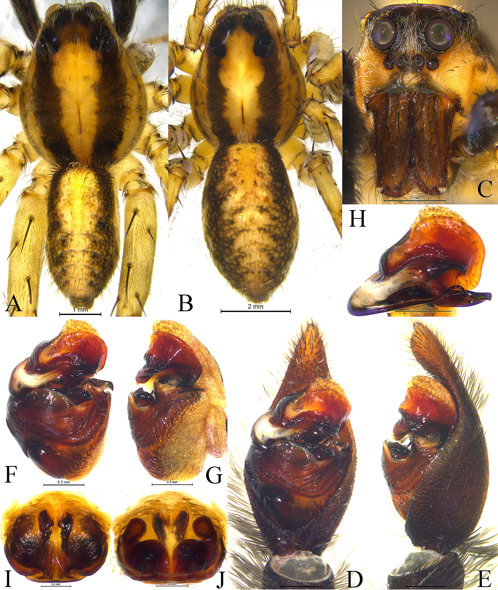
FIGURE 4. Pardosa tuberosa sp. nov., male holotype (A, C, D, E) and female paratype (B, I, J) from the area near Mannasan Bridge, male paratype (F, G, H) from Manxie tunnel. A. Male habitus, dorsal view; B. Female habitus, dorsal view; C. Male eyes and chelicerae, front view; D. Left male palp, ventral view; E. Same, retrolateral view; F. Bulb, ventral view; G. Same, retrolateral view; H. Apical part of bulb, ventral view; I. Epigyne, ventral view; J. Same, dorsal view.
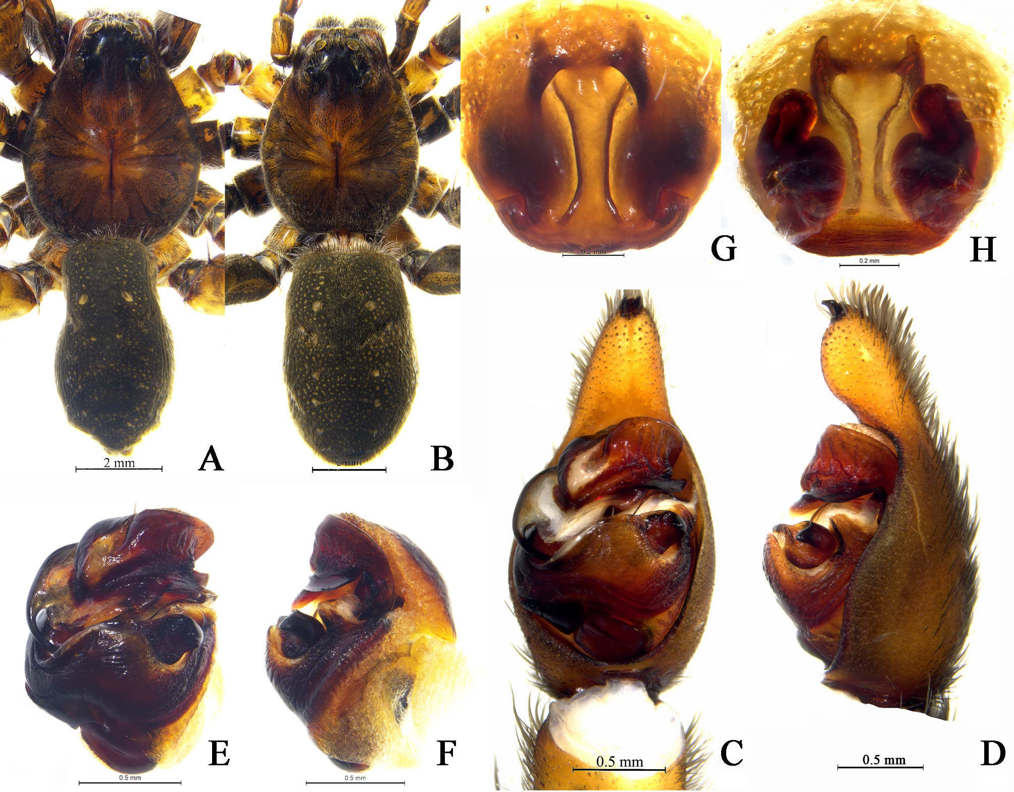
FIGURE 8. Pardosa chapini (Fox, 1935), male and female from Zhaojue Country, Sichuan Province, China. A. Male habitus, dorsal view; B. Female habitus, dorsal view; C. Left male palp, ventral view; D. Same, retrolateral view; E. Bulb, ventral view; F. Same, retrolateral view; G. Epigyne, ventral view; H. Same, dorsal view.
No known copyright restrictions apply. See Agosti, D., Egloff, W., 2009. Taxonomic information exchange and copyright: the Plazi approach. BMC Research Notes 2009, 2:53 for further explanation.
1 (by plazi, 2016-04-17 10:49:14)
2 (by ImsDioSync, 2017-01-18 20:52:41)
3 (by ImsDioSync, 2017-01-18 20:53:20)
4 (by ImsDioSync, 2017-06-25 00:56:42)
5 (by ExternalLinkService, 2019-09-26 14:05:38)
6 (by ExternalLinkService, 2022-01-30 06:14:32)
7 (by ExternalLinkService, 2022-02-18 09:14:50)
8 (by plazi, 2023-10-26 22:31:54)