
Ophion kevoensis Jussila, 1965
|
publication ID |
https://doi.org/10.5852/ejt.2019.550 |
|
publication LSID |
lsid:zoobank.org:pub:F8707194-B55E-48CA-8FE0-4CD0D023C217 |
|
DOI |
https://doi.org/10.5281/zenodo.3477039 |
|
persistent identifier |
https://treatment.plazi.org/id/A270EE7E-FC3B-FFA2-F363-A89635E6F817 |
|
treatment provided by |
Plazi |
|
scientific name |
Ophion kevoensis Jussila, 1965 |
| status |
|
Ophion kevoensis Jussila, 1965 View in CoL
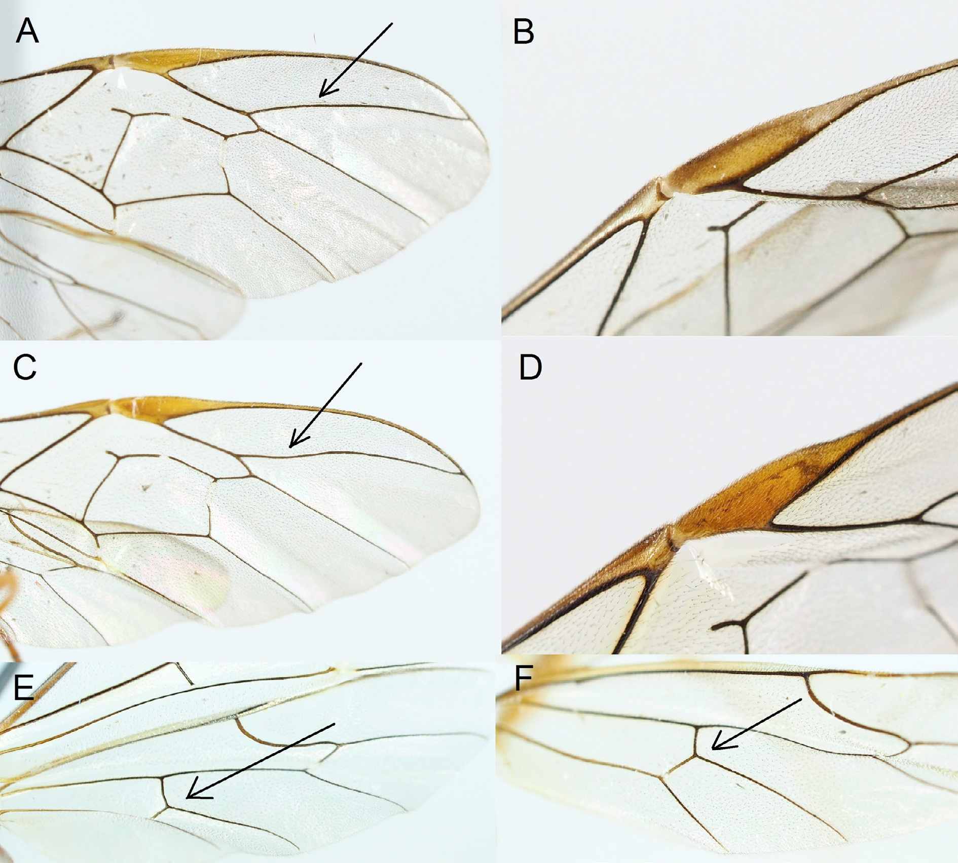
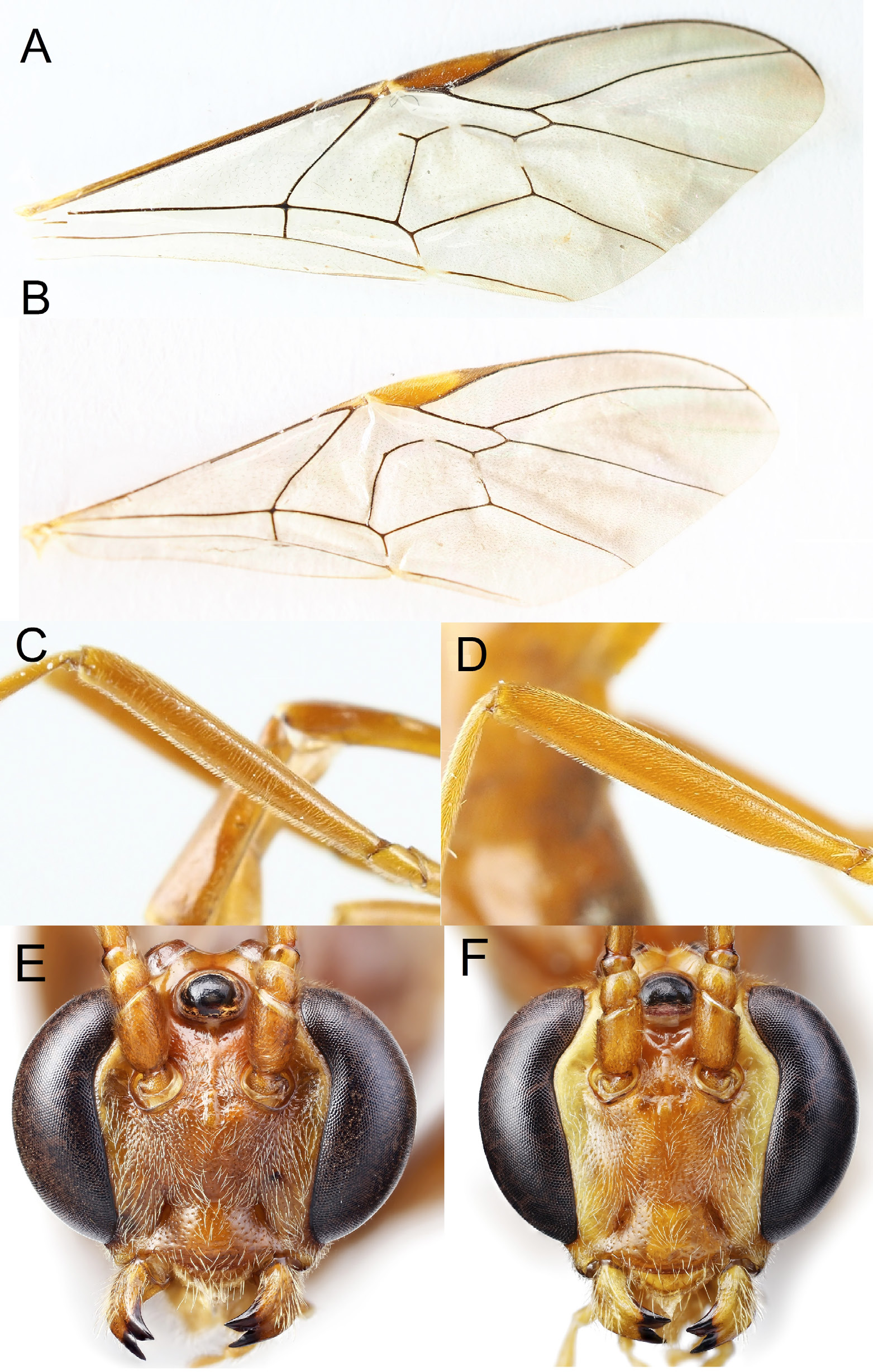
Figs 13D View Fig , 16E View Fig , 21E View Fig
Ophion kevoensis Jussila, 1965: 97–98 View in CoL .
Material examined
Holotype, ♂, and allotype, ♀ ( RJC); 95 ♀♀, 46 ♂♂ ( Sweden); 4 ♀♀ ( Finland); 8 ♀♀ ( Norway).
Diagnosis
Fore wing length 15–17 mm. Antenna with 52–57 flagellomeres. First flagellomere about 3.5–4.0 times as long as wide. Central flagellomeres about 1.5–1.6 times as long as wide. Subapical flagellomeres approximately 1.5 times as long as wide. Temple buccate, in lateral view 0.6–0.8 times as long as compound eye. Head in dorsal view with gap between compound eye and lateral ocellus about 0.2–0.3 times the diameter of the ocellus. Malar space in both sexes, especially in the male, slightly longer than in O. slaviceki ( Fig. 21E View Fig ), in female about 0.3 times as long as mandibular base and 0.5–0.7 in males. Face wide ( Fig. 16E View Fig ) below antennal sockets densely and deeply punctate. Gap between mandibles obtuse or right angled, with internal angles present. Wing membrane usually yellowish. Ramellus long, 0.3 times the width of the discosubmarginal cell. Radius sinuous. Structure of mesopleuron weakly shagreened with distinct punctures. Interstices between punctures about equal to their diameter. Epicnemial carina, in antero-ventral view, with pleurosternal angles obviously anterior to sternal angles. Pleurosternal angles rounded, obtuse to right angled (as in Fig. 9E View Fig ). Scutellum with lateral carinae present in basal 0.5–0.6. Anterior transverse carina of propodeum present, quite strong. Posterior transverse carina only present laterally, widely interrupted centrally. Petiolar carina and longitudinal carina delimiting area superomedia weak. Sclerotised part of first sternite ending level or slightly posterior to spiracle. Hind trochantellus shorter than wide in dorsal view. Legs normal, with hind femur about 6.0–6.5 times as long as wide. Hind trochantellus shorter than wide in dorsal view. Inner spur of hind tibia about 0.3–0.4 times as long as hind metatarsus.
Colour
Mesosoma usually brownish testaceous. Mandibular teeth black. Head with inner and outer orbits reddish-yellow, distinctly darker than in O. slaviceki . Apical flagellomeres in male slightly infuscate. Pterostigma evenly testaceous ( Fig. 13D View Fig ). Ovipositor sheath of the same colour as posterior segments of metasoma.
DNA barcode
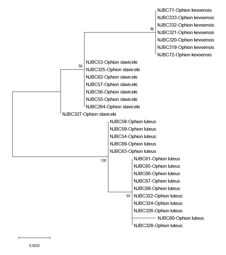
The DNA barcode sequences of seven Swedish specimens of Ophion kevoensis are available at the BOLD systems database (www.boldsystems.org, BIN. BOLD: ACF9514. Specimen codes: STI-NJBC: 71–72, 319–321, 332–333).
Ecology
The species occurs in semi-open coniferous forests with Vaccinium L. during August–October in Central and Northern Sweden.
Distribution in Sweden
Widespread and locally very abundant in subalpine taiga forests where it is the dominant Ophion species.
Remarks
A rather variable species. Closely related to O. slaviceki , but easily distinguished from that species by the partly carinated scutellum, the generally longer ramellus, the right angled mandibular gape, the generally shorter hind trochantellus, the usually yellowish wing membrane, the longer malar space, especially in the male, and the usually darker colour. Also very similar to O. autumnalis Johansson sp. nov., but on average larger and with the pterostigma evenly testaceous ( Fig. 13D View Fig ). Sometimes the carinae of the propodeum can be quite distinct and the sclerotised part of first sternite end posterior to spiracle in which case the species can be mistaken for Ophion arenarius . Ophion kevoensis is distinguished from that species by the more buccate head with a distinct gap between the lateral ocellus and the inner margin of the compound eye. The barcoded specimens form a group that is closely related to O. slaviceki ( Fig. 3 View Fig ). The allotype female ( Jussila 1966) is smaller, has a narrow face and the ovipositor sheath black, contrasting with the posterior metasomal segments. The specimen probably represents a different species, most likely Ophion sylvestris Johansson sp. nov. (see further comments under Discussion-‘additional comments on type material’). It is important to note that the interpretation of this species in this study is based on detailed drawings made by the second author when he gained access to the type material in the 1970-s. For the present paper, the types were unavailable for detailed examination as they were on loan for a study of Ophion species in Finland (Gergely Várkonyi, Finnish Environment Institute, pers. com.).


No known copyright restrictions apply. See Agosti, D., Egloff, W., 2009. Taxonomic information exchange and copyright: the Plazi approach. BMC Research Notes 2009, 2:53 for further explanation.
|
Kingdom |
|
|
Phylum |
|
|
Class |
|
|
Order |
|
|
Family |
|
|
SubFamily |
Ophioninae |
|
Genus |
Ophion kevoensis Jussila, 1965
| Johansson, Niklas & Cederberg, Björn 2019 |
Ophion kevoensis
| Jussila 1965: 97 |