
Homoneura (Homoneura) simigrandis, Shi, Li & Yang, Ding, 2014
|
publication ID |
https://doi.org/10.11646/zootaxa.3890.1.1 |
|
publication LSID |
lsid:zoobank.org:pub:F74FF49D-BA75-441F-B6E3-38D5DCA92048 |
|
DOI |
https://doi.org/10.5281/zenodo.5732250 |
|
persistent identifier |
https://treatment.plazi.org/id/F871DC59-53F9-41C0-8A63-DAC96C1041A9 |
|
taxon LSID |
lsid:zoobank.org:act:F871DC59-53F9-41C0-8A63-DAC96C1041A9 |
|
treatment provided by |
Plazi (2016-04-18 14:31:10, last updated 2024-11-24 23:21:03) |
|
scientific name |
Homoneura (Homoneura) simigrandis |
| status |
sp. nov. |
Homoneura (Homoneura) simigrandis View in CoL sp. nov.
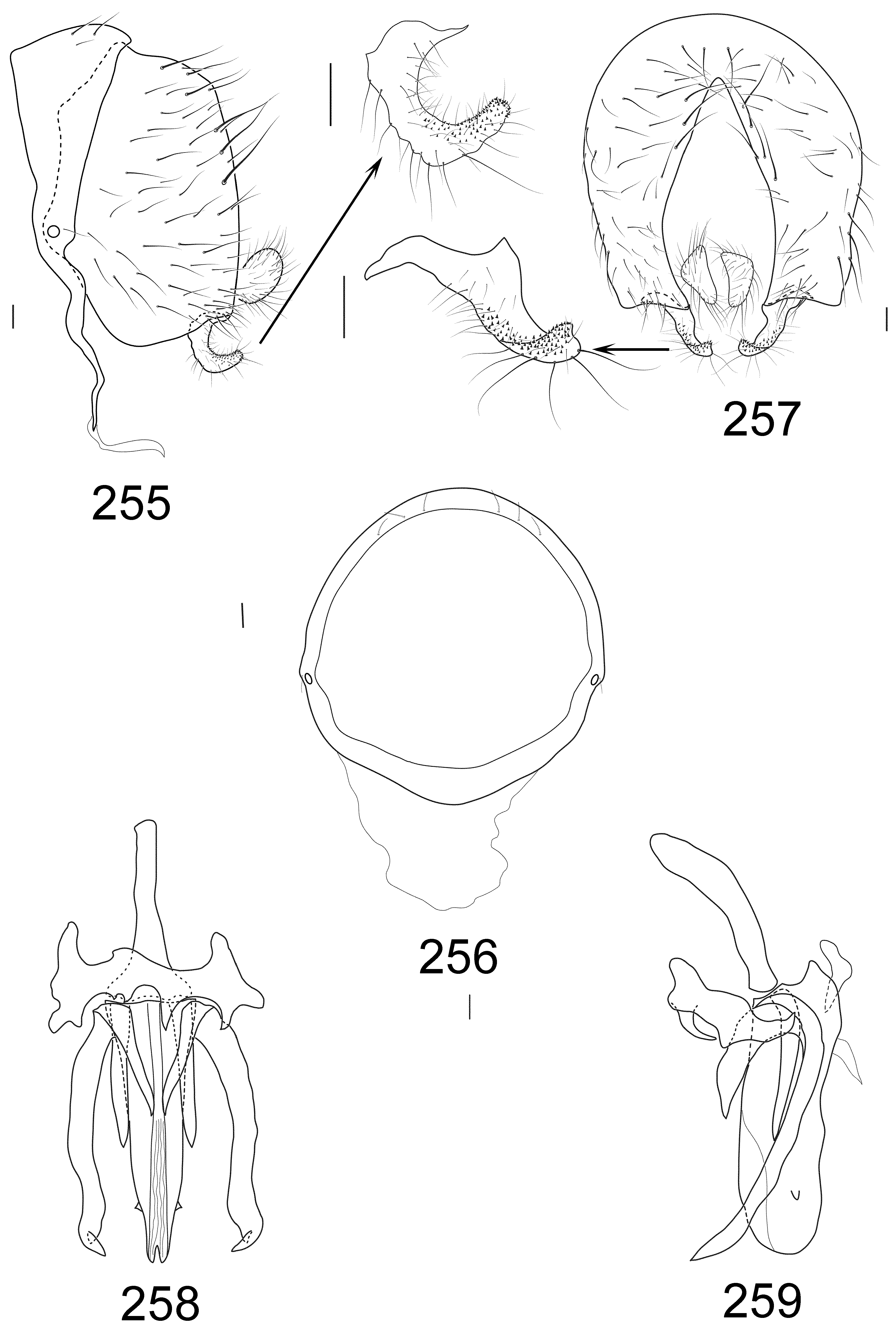
( Figs 249–259 View FIGURES 249 – 254 View FIGURES 255 – 259 )
Diagnosis. Wing with basal edges of brown apical spots on R2+3, R4+5 and M1 behind vertical level of brown stripe on dm-cu, forming ladder-like slope; a small round spot on R4+5, nearly middle point of distance between r-m and dm-cu; brown stripe over dm-cu with brown spot on upper margin larger than that on lower margin; subcostal cell pale brown. Male genitalia: reverse U-shaped pregonite with two arms asymmetrical distinctly and postgonite with a short process; aedeagus with a pair of lateral teeth subapically in ventral view
Description. MALE. Body length 6.7 mm, wing length 6.9 mm.
Head ( Figs 250–251 View FIGURES 249 – 254 ) yellow. Frons brownish yellow except for pale yellow anterior marign, as long as wide; ocellar triangle yellow; oc developed, longer than anterior or; anterior or slightly shorter than posterior or; gena about 1/5 height of eye. Antenna yellow, 1st flagellomere pale grayish brown on apical 1/3 (only one present, another one broken); 1st flagellomere 2.0 times longer than height; arista dark except for brownish basally, ray short plumose with longest ray slightly shorter than height of 1st flagellomere. Proboscis and palpus yellow.
Thorax ( Fig. 252 View FIGURES 249 – 254 ) yellow. Mesonotum with 0+3 dc (anteriormost dc distinctly behind transverse suture), acr in irregular 8 rows, prsc as long as anteriormost dc. Legs yellow, tarsomeres 3–5 pale brown. Fore femur with 4 strong pv, 8 pd, and ctenidium with 12 short seta; fore tibia with 1 long preapical ad and 1 short apv. Mid femur with 4 a and 2 app; mid tibia with 1 strong preapical ad and 3 strong apv. Hind femur with 3 av (on apical 1/4) and 1 preapical ad; hind tibia with 1 preapical ad and 1 short apv. Wing ( Fig. 254 View FIGURES 249 – 254 ) pale yellow, basal edges of brown apical spots on R2+3, R4+5 and M1 behind vertical level of brown stripe on dm-cu, forming ladder-like slope; a small round spot on R4+5, nearly middle point of distance between r-m and dm-cu; brown apical spots on R2+3 and R4+5 confluent, apical spot on M1 separated from that on R4+5; brown stripe over dm-cu with brown spot on upper margin larger than that on lower margin; subcostal cell pale brown; costa with 2nd (between R1 and R2+3), 3rd (between R2+3 and R4+5) and 4th (between R4+5 and M1) sections in proportion of 5.2:1.3:1; r-m before middle of discal cell; ultimate and penultimate sections of M 1 in proportion of 1:1.6; ultimate section of CuA1 about 1/7 of penultimate. Halter yellow.
Abdomen ( Fig. 253 View FIGURES 249 – 254 ) yellow. Male genitalia ( Figs 255–259 View FIGURES 255 – 259 ): syntergosternite circular with dorsal setulae; epandrium broad, with 5 pairs of long dorsal setae in ventral view; surstylus curved with long setulae and many tiny teeth in lateral view; hypandrium nearly H–shaped with small acute ventral processes in different length, and hypandrial apodeme short; reverse U-shaped pregonite with two arms asymmetrical distinctly and postgonite with a short process; aedeagus with a pair of lateral teeth subapically in ventral view; aedeagal apodeme long.
Material examined. 1 ♂ ( USNM), CHINA, Kwantung, Yim Na San E., 10. VI. 1936, A.L.Melander collection 1961.
Distribution. China (Guangdong).
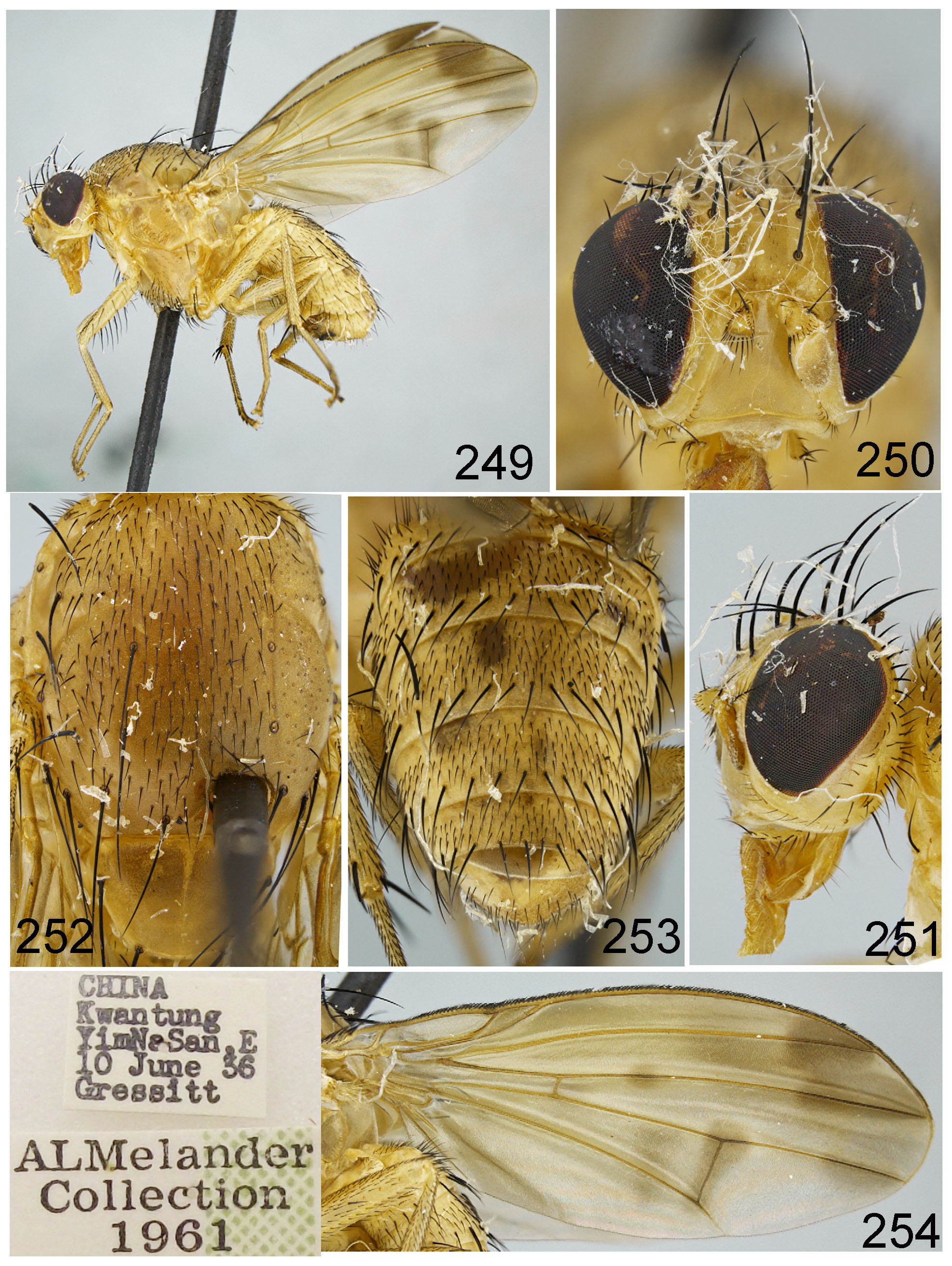
FIGURES 249 – 254. Homoneura (Homoneura) simigrandis sp. nov. Male. 249. body, lateral view; 250, 251. head, anterior view; 252. thorax, dorsal view; 253. abdomen, dorsal view; 254. wing.
| USNM |
Smithsonian Institution, National Museum of Natural History |
No known copyright restrictions apply. See Agosti, D., Egloff, W., 2009. Taxonomic information exchange and copyright: the Plazi approach. BMC Research Notes 2009, 2:53 for further explanation.
|
Kingdom |
|
|
Phylum |
|
|
Class |
|
|
Order |
|
|
Family |
|
|
Genus |
1 (by plazi, 2016-04-18 14:31:10)
2 (by ImsDioSync, 2017-01-18 13:59:35)
3 (by ImsDioSync, 2017-01-18 14:04:49)
4 (by ExternalLinkService, 2019-09-26 13:21:11)
5 (by ExternalLinkService, 2021-11-27 19:41:14)
6 (by ExternalLinkService, 2021-11-27 19:57:26)
7 (by ExternalLinkService, 2021-11-27 20:24:17)
8 (by plazi, 2023-10-29 06:40:08)