
Adlafia ossiformis, Alibert & Hamilton & Pienitz & Antoniades, 2023
|
publication ID |
https://doi.org/10.5252/cryptogamie-algologie2023v44a9 |
|
DOI |
https://doi.org/10.5281/zenodo.10392936 |
|
persistent identifier |
https://treatment.plazi.org/id/AB688783-2B3E-FFEA-FC12-F8D7FED34D54 |
|
treatment provided by |
Plazi (2023-12-14 10:21:37, last updated 2024-11-27 00:55:56) |
|
scientific name |
Adlafia ossiformis |
| status |
sp. nov. |
Adlafia ossiformis sp. nov.
( Figs 10 View FIG AH-AT; 13 View FIG A-H)
HOLOTYPE. — Canada. Québec, Lake 22AA, 56°33’24”N, 76°28’47”W, 128 m a.s.l., 22.VIII.2015, D. Antoniades (microscope slide designated as the holotype, holo-, CANA [ CANA 129481 ]) GoogleMaps .
ISOTYPE. — Canada. Québec, Lake 22AA, 56°33’24”N, 76°28’47”W, 128 m a.s.l., 22.VIII.2015, D. Antoniades (iso-, ANSP [ ANSP-GC68069 ]) GoogleMaps .
TYPE LOCALITY. — Canada. Québec, Lake GoogleMaps 22AA, 56°33’24”N, 76°28’47”W, 128 m a.s.l.
ETYMOLOGY. — The epithet ‘ossiformis’ is Latin, describing this species’ bone-shaped valves.
ECOLOGY AND DISTRIBUTION. — This species was found only in 22-AA lake of the study, where it represented 4% of the diatom assemblage. Lake 22-AA had the highest TP concentration in the dataset (23.4 µg/L).
REGISTRATION. — http://phycobank.org/103920.
DESCRIPTION
Frustules rectangular and narrow in girdle view.Valves elliptic at mid-valve with broad capitate ends. Valve dimensions (n =20): length 13.5-20.5 µm, width 4.5-5.5 µm and 30-35 striae in 10 µm. External valve face flat, margin rounded. Axial area slightly curved to linear and narrow. Central area round to elliptic,>1/2 width of valve, with 5-7 marginal striae between proximal raphe endings. Raphe linear, with no kink-like irregularity halfway between mid-valve and apex ( Fig. 13A View FIG ); externally, central raphe fissures widely spaced with small indistinct ends bent to one side ( Fig. 13C View FIG ). Terminal raphe fissures curved down onto upper region of mantle ( Fig. 13G View FIG ). A weakly formed groove present parallel to raphe on mantle up to valve face. Internally, raphe on small sternum, straight, with no kink-like irregularity ( Fig. 13B View FIG ). Proximal raphe fissures bent and deflected on a raised nodule ( Fig. 13D View FIG ). One surface depression present on central nodule ( Fig. 13D, E View FIG ). Terminal fissures end on small elongated helictoglossae, isolated from apex mantle ( Fig. 13H View FIG ). Hyaline area of terminal nodule more developed on secondary side of valve. Striae continuous, straight to weakly arched from valve face to mantle, more widely spaced around central area. Voigt faults weak to indistinct on secondary side of valve 2/3 of distance between mid-valve and apex. Striae continuous around base of apices. Areolae round to rectangular, 4-6 per stria. Areolae covered with a fine poroid velum with 8-9 circular rows of small pores around outside edge and linear rows on inner area. Internally, areolae recessed between virgae.
Adlafia ossiformis sp. nov. has a shape that is easily confused in LM with Psammothidium ventralis (Krasske) Bukhtiyarova & Round and Sellaphora guyanensis Metzeltin & Lange-Bertalot , but careful examination of both valves will make species differentiation possible.
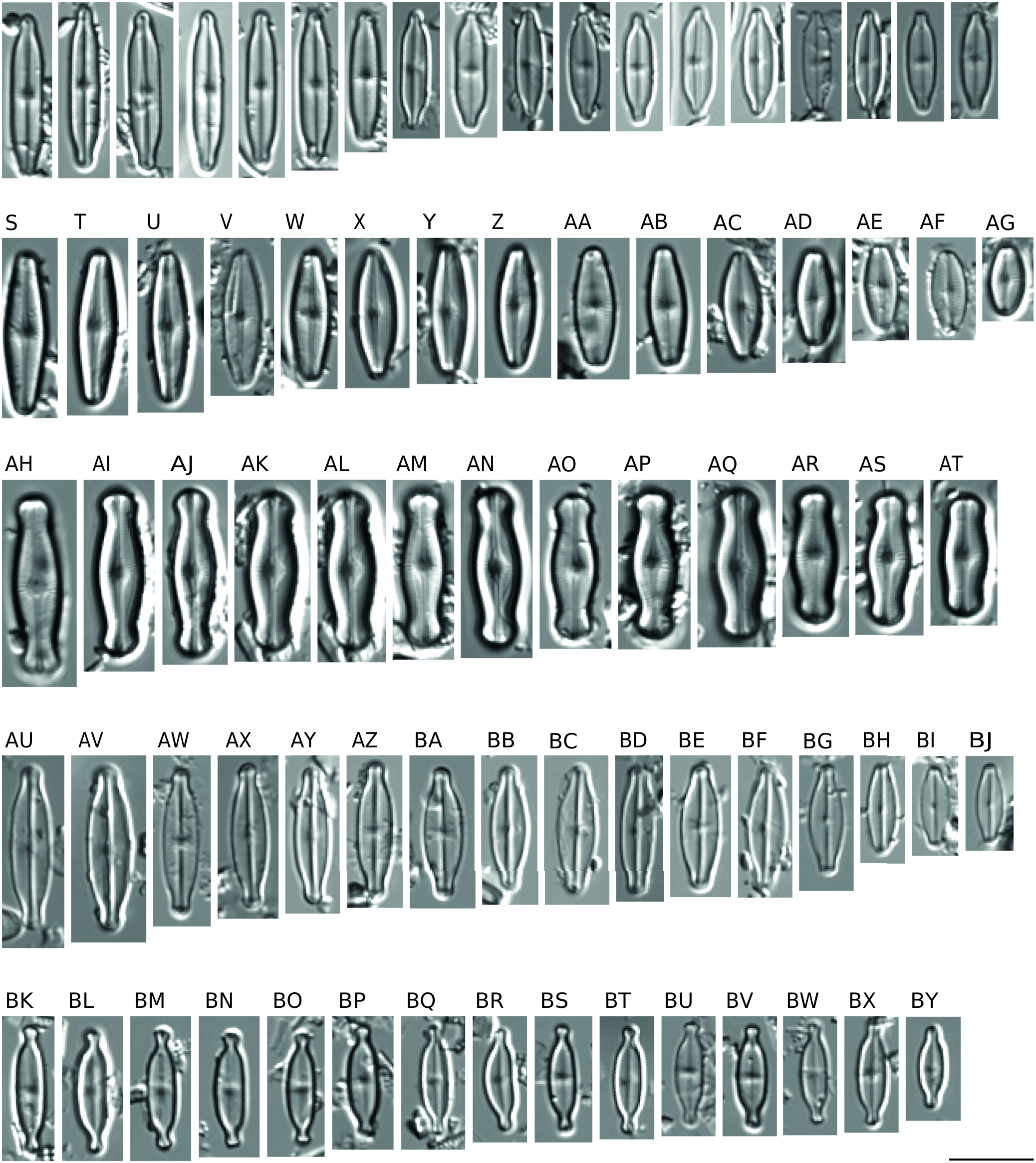
FIG. 10. — Light micrograph diminution series of Adlafia Moser,Lange-Bertalot & Metzeltin,Sellaphora Mereschkowski and Nupela Vyverman & Compère species: A-R, Adlafia bryophila (J.B.Petersen) Lange-Bertalot; S-AG, Adlafia umiujaqensis sp. nov.; AH-AT, Adlafia ossiformis sp. nov.; AU-BJ, Sellaphora vincentiana sp. nov.; BK-BY, Nupela tenuicephala (Hustedt) Lange-Bertalot. Scale bar: 10 µm.
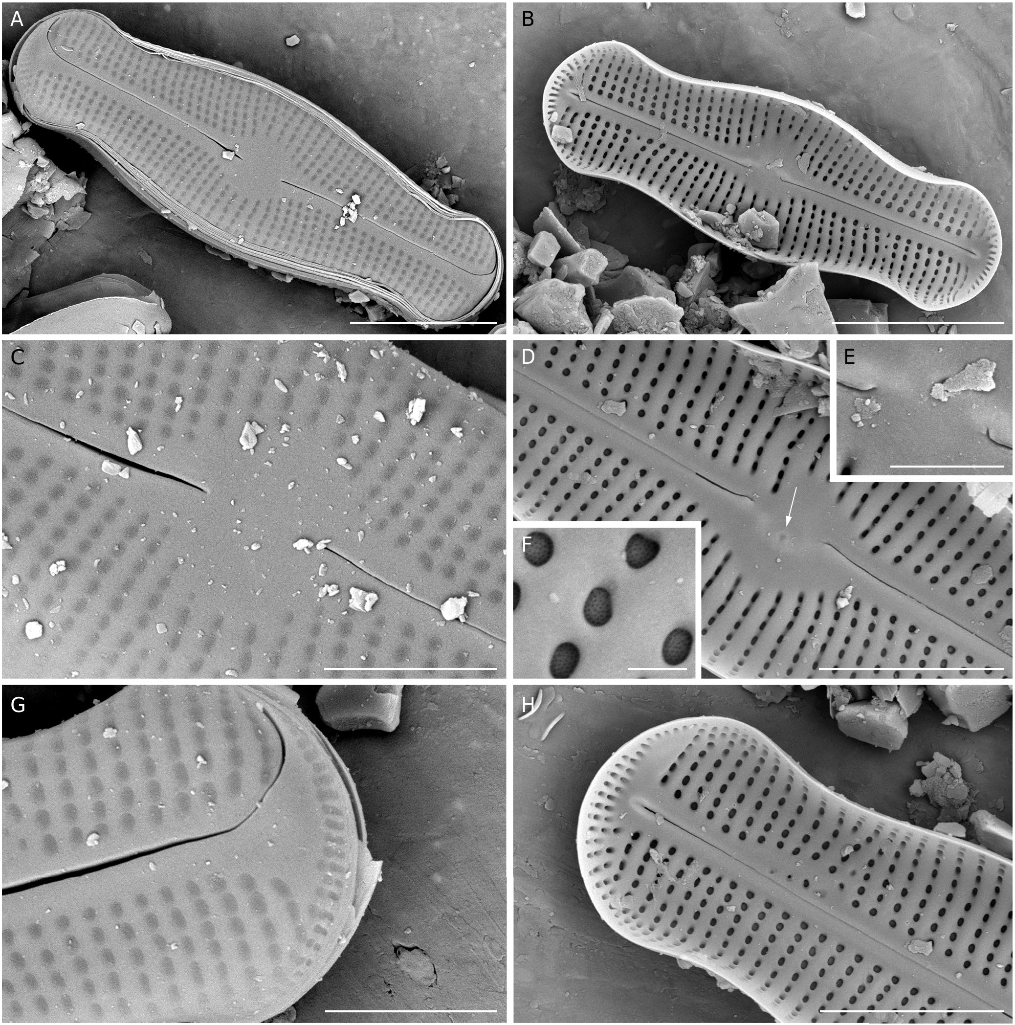
FIG. 13. — SEM images of Adlafia ossiformis sp. nov.:A, external view of the complete valve; B, internal view of the complete valve; C, external view of the central area showing small proximal raphe fissures and multiseriate poroid covers over the areolae; D, internal view of the central area with curved fissures and a ghostareolae depression; E, magnification of the central area; F, magnification of areolae with recessed poroid volae; G, external view of the apex. Fissure bent down onto the mantle; H, internal view of the apex. The central nodule is elevated and an isolated pore (arrow) may be present. Scale bars: A, B, 5 µm; C, G, 2 µm; D, H, 3 µm; E, 1 µm; F, 300 nm.
| ANSP |
Academy of Natural Sciences of Philadelphia |
No known copyright restrictions apply. See Agosti, D., Egloff, W., 2009. Taxonomic information exchange and copyright: the Plazi approach. BMC Research Notes 2009, 2:53 for further explanation.
|
Kingdom |
|
|
Phylum |
|
|
Class |
|
|
Order |
|
|
Family |
|
|
Genus |