
Phyllocnistis tropaeolicola Kawahara, Nishida & Davis, 2009
|
publication ID |
https://doi.org/10.3897/zookeys.27.250 |
|
publication LSID |
lsid:zoobank.org:pub:C6AA8595-6A57-4ACD-B0A1-3AE36F7C8701 |
|
persistent identifier |
https://treatment.plazi.org/id/E766981C-D9EC-48DF-9C9D-D02EC2AC9744 |
|
taxon LSID |
lsid:zoobank.org:act:E766981C-D9EC-48DF-9C9D-D02EC2AC9744 |
|
treatment provided by |
Plazi (2020-04-27 06:56:54, last updated by Admin 2020-04-28 00:34:11) |
|
scientific name |
Phyllocnistis tropaeolicola Kawahara, Nishida & Davis |
| status |
sp. n. |
Phyllocnistis tropaeolicola Kawahara, Nishida & Davis , sp. n.
urn:lsid:zoobank.org:act:E766981C-D9EC-48DF-9C9D-D02EC2AC9744
Diagnosis ( Table 1). Phyllocnistis tropaeolicola differs from P. drimiphaga and P. maxberryi in its larger size, having a slender longitudinal fascia, valva that are ̴2.4× the length of the vinculum, and a single, band-shaped signa. The pupa of P. tropaeolicola has conical frontal processes and dorsal abdominal spines on each segment are arranged in a V.
Adult ( Fig. 2C View Figure 2 ). Forewing length 2.6–5.0 mm. Head. Vestiture silvery white, completely covered with smooth, broad, scales slightly overlapping anterior margin of eyes. Antenna ̴ equal to length of forewing, scape and pedicel enlarged laterally and covered in long silvery scales, a single row of fine short scales completely encircling each flagellomere. Labial palpus long, slender, ̴ 1.0 mm. Thorax. Forewing silvery white; with a slender, dark-brown, longitudinal fascia extending 2/3 length of wing to meet distally at junction of brown, costal and transverse fasciae; costal fascia slender and strongly oblique with dark-brown border; transverse fascia V-shaped, with a dark-brown border; apical to subapical area pale yellowish orange with a small black spot; three slender, dark-brown costal strigulae, three slender dark-brown apical strigulae, and one faint brown tornal strigula arising from black apical spot; fringe along tornal margin white with a dark-brown basal band of broad scales. Hindwing mostly white except for a band of pale brown scales extending length of costal margin. Legs similar to P. drimiphaga , silvery white except dark brown over dorsal surface of femur, tibia and tarsus of foreleg. Abdomen. Length ̴ 2.0 mm, mostly brownish gray dorsally, silvery white ventrally. Coremata similar to P. drimiphaga . Male genitalia ( Figs 6 View Figure 6 A–C). Similar to P. drimiphaga except valva relatively longer and more slender, ̴ 2.4× the length of vinculum, nearly straight, with ventral lobe of apex slightly re-curved dorsad ( Fig. 6A View Figure 6 ). Genitalia slide USNM 33281. Female genitalia ( Figs 6D, E View Figure 6 ). Oviscapt greatly reduced as in P. drimiphaga ; ductus bursae completely membranous, slender, elongate, ̴ 8.5× length of papillae anales and terminating at posterior end of corpus bursae; corpus bursae ̴ 0.6× length of ductus bursae; a single elongate signum present as a narrow band partially encircling middle of corpus bursae; signum with 2 acute, flattened spines projecting inwards from band; length of spines slightly more than width of signa; ductus seminalis extremely slender, elongate, ̴ 2.4 × length of corpus bursae and arising from near middle of corpus bursae. Genitalia slide USNM 33282, 33285, 33288.
Larva ( Figs 12A, C–F). Young sap-feeding larva translucent yellow ( Fig. 12A). Mature sap-feeding larva ̴ 7.5 mm long, translucent yellow, head capsule translucent pale brown, prothoracic shield dark brown ( Figs 12 C–). Cocoon-spinning larva whitish yellow, head capsule pale gray brown; ̴ 6.5 mm long ( Fig. 12F).
Pupa ( Figs 10, 12H). Brown, length up to ̴ 5 mm; diameter ̴ 1.0 mm. Vertex with a short, stout, process (cocoon-cutter) flanked by two, flattened, slightly longer processes ( Figs 9A, B, D, E View Figure 9 ) and two pairs of short setae ( Fig. 9C View Figure 9 ). Dorsum of A2–A7 with a pair of laterally curved, large spines in between which is a concentration of smaller spines, arranged in a triangular, V-shaped pattern ( Figs 9F, G View Figure 9 ); each segment with a pair of long, lateral, sensory setae ( Fig. 9L View Figure 9 ) that are shortest on A9–10 ( Figs 9J, K View Figure 9 ). A10 with a pair of slightly divergent processes from caudal apex ( Figs 9I, J View Figure 9 ).
Types. Holotype ( Fig. 2C View Figure 2 ): ♂, Costa Rica: Prov. Cartago, Cerro de la Muerte, Villa Mills, 3100 m, 13 Mar 2003 (adult emergence), host Tropaeolum emarginatum , col./ rear Kenji Nishida, mine with pupal fold collected 6 Mar 2003 ( USNM) . Paratypes: Immatures: 1 prepupa, 1 pupa ( USNM 34036 ), Villa Mills , Georgina , 9°33'30"N, 83°43'25.8"W, 3103 m, 12 Sep 2008, K. Nishida, host Tropaeolum emarginatum GoogleMaps . Adults: same locality as holotype, 6♂, 4♀: ♂ slide USNM 33281 , ♀ slide USNM 33285 GoogleMaps ; 2♂, 2♀ ( USNM 33280 About USNM , 33282 About USNM ) with adult emergence 11 Mar 2003 ; 1♂, with adult emergence 15 Mar 2003. 1♀ adult paratype at INBio and UCR, the remaining paratypes at USNM .
Life history ( Fig. 12). Mines of P. tropaeolicola were readily found on plants growing along the Pan-American Highway ( Fig. 1H). Most mines occurred on full-grown new leaves ( Figs 12B, C) but some were found on developing leaves ( Fig. 12A). Thirteen had a single mine, two leaves had two, and one had three. All mines were found on the adaxial side, and the late sap-feeding instar fed on the mesophyll ( Fig. 12E).
The mine characteristically begins as a narrow, irregular serpentine gallery ( Fig. 12B) that widens as it extends along or near the leaf margin ( Figs 12B, C). It is relatively narrow, pale green to white with a less conspicuous dark green median frass line. Pupal cocoon folds were ̴ 5.5 mm long and were found near the leaf margin ( Figs 12B, G). Adults emerged 5–9 days after pupal cocoon folds were collected.
We found mines of an unidentified fungus gnat ( Diptera : Mycetophilidae ) at same site on the same plant. The mines, which usually occur several on a single leaf, are irregularly shaped blotch mines with dark-green frass scattered randomly within. The fly larva causes curling, drying, necrosis, and yellowing of the leaves, and was more abundant than P. tropaeolicola mines. Several leaves were infested with both mycetophilid and P. tropaeolicola larvae.
Host. Tropaeolum emarginatum Turcz ( Tropaeolaceae ) ( Fig. 1I). Tropaeolum , the only genus recognized in Tropaeolaceae , is Neotropical and contains approximately 90 species, many of which are found in Andean cloud forests ( Gentry 1996). Four species occur in Costa Rica, and T. emarginatum is present on both the Atlantic and Pacific slopes between 700 and 3200 m ( Alfaro-Vindas 2003; INBio 2009). Outside Costa Rica, T. emarginatum has been recorded from Chiapas, Mexico to Cotopaxi, Ecuador ( Missouri Botanical Garden 2009). The tenuous, soft, and succulent vines of T. emarginatum are usually found in forest edges and disturbed areas, and the flowers are red to yellow orange ( Alfaro-Vindas 2003; Gentry 1996). Most of the leaves are between 5 and 8 cm wide ( KN, pers. obs.).
Distribution. Known only from the type locality, Cerro de la Muerte, Villa Mills, at 3100 m elevation in the Cordillera de Talamanca.
Etymology. The species name, tropaeolicola , is formed from its host plant genus name, Tropaeolum , and the Latin word cola, meaning “inhabitant”.
Alfaro-Vindas E (2003) Plantas comunes del Parque Nacional Chirripo = Common plants of Chirripo National Park - Costa Rica. Instituto Nacional de Biodiversidad, Santo Domingo de Heredia, 268 pp.
Gentry AH (1996) A field guide to the families and genera of woody plants of northwest south America (Colombia, Ecuador, Peru) with supplementary notes on herbaceous taxa. University of Chicago Press, Chicago, x + 895 pp.
Missouri Botanical Garden (2009) Tropicos. org. Missouri Botanical Garden. Available from http: // www. tropicos. org / [accessed 19 April 2009].
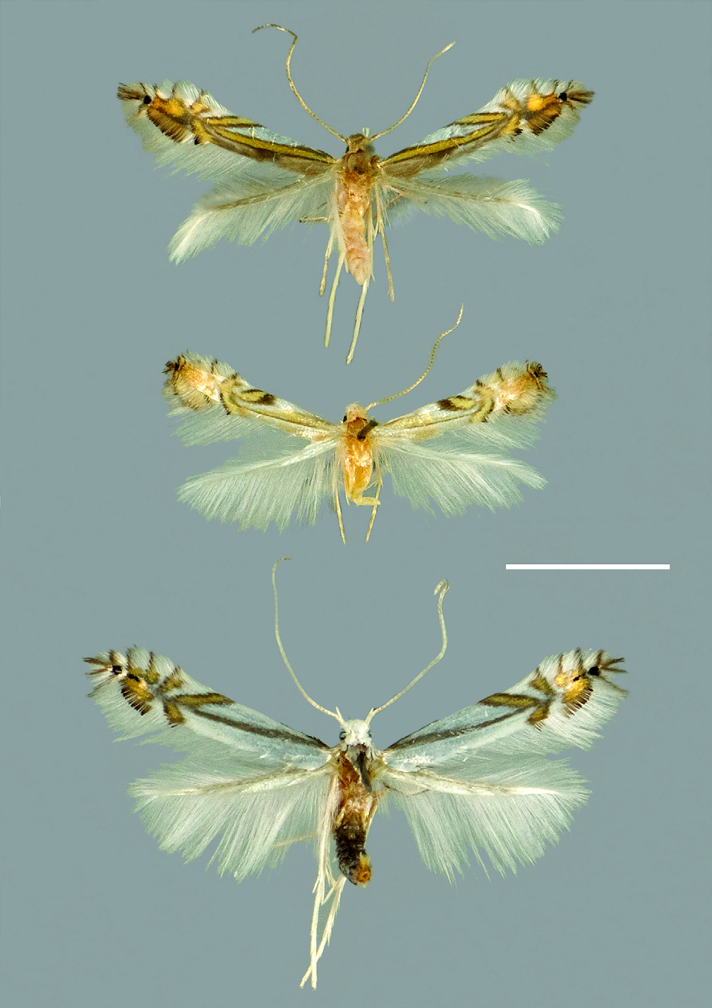
Figure 2. Adults of three new Phyllocnistis species from Costa Rica. A Phyllocnistis drimiphaga sp. n., holotype female B P. maxberryi sp. n., holotype female (abdomen removed for dissection) C P. tropaeolicola sp. n., holotype male.
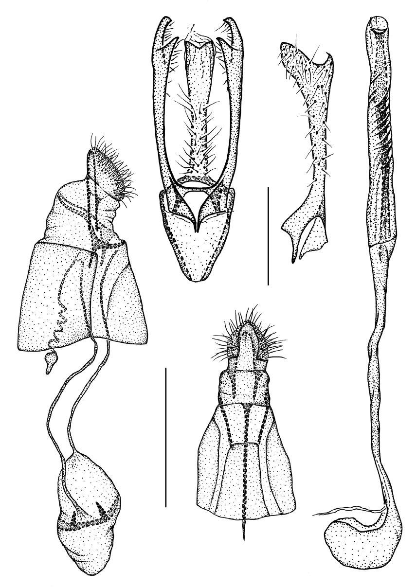
Figure 6. Phyllocnistis tropaeolicola sp. n., genitalia. A Male, ventral view B right valva, mesal view C aedeagus D female, lateral view E ventral view of terminal segments. (Scale bar 0.5 mm except for figure B, 0.25 mm.)

Figure 9. Phyllocnistis tropaeolicola sp. n., pupa. A Ventral view of head B ventral view of cocoon-cutter C frons D lateral view of head E lateral view of cocoon-cutter F dorsal view of fourth abdominal tergum G spines on fourth abdominal tergum H lateral view of spines on fourth abdominal tergum I view of abdominal tip Į dorsal view of A9–10 K lateral seta on A9–10 L lateral seta on seventh abdominal tergum. Scale bars 100 µm.
No known copyright restrictions apply. See Agosti, D., Egloff, W., 2009. Taxonomic information exchange and copyright: the Plazi approach. BMC Research Notes 2009, 2:53 for further explanation.
|
Kingdom |
|
|
Phylum |
|
|
Class |
|
|
Order |
|
|
Family |
|
|
Genus |
1 (by plazi, 2020-04-27 06:56:54)
2 (by ExternalLinkService, 2020-04-28 00:44:22)
3 (by ExternalLinkService, 2020-05-06 02:59:39)
4 (by ExternalLinkService, 2021-11-09 13:44:10)
5 (by ExternalLinkService, 2021-11-10 00:13:56)
6 (by plazi, 2023-10-31 09:03:20)