
Astraeus ( Depollus ) aberrans van de Poll, 1886
|
publication ID |
https://doi.org/10.5281/zenodo.5372066 |
|
publication LSID |
lsid:zoobank.org:pub:F5F00646-B3F6-47F8-9CD4-20B6B448BEEC |
|
DOI |
https://doi.org/10.5281/zenodo.5332098 |
|
persistent identifier |
https://treatment.plazi.org/id/03F587D5-E622-FF94-FE80-FAEDFD0EFBD8 |
|
treatment provided by |
Marcus |
|
scientific name |
Astraeus ( Depollus ) aberrans van de Poll, 1886 |
| status |
|
Astraeus ( Depollus) aberrans van de Poll, 1886
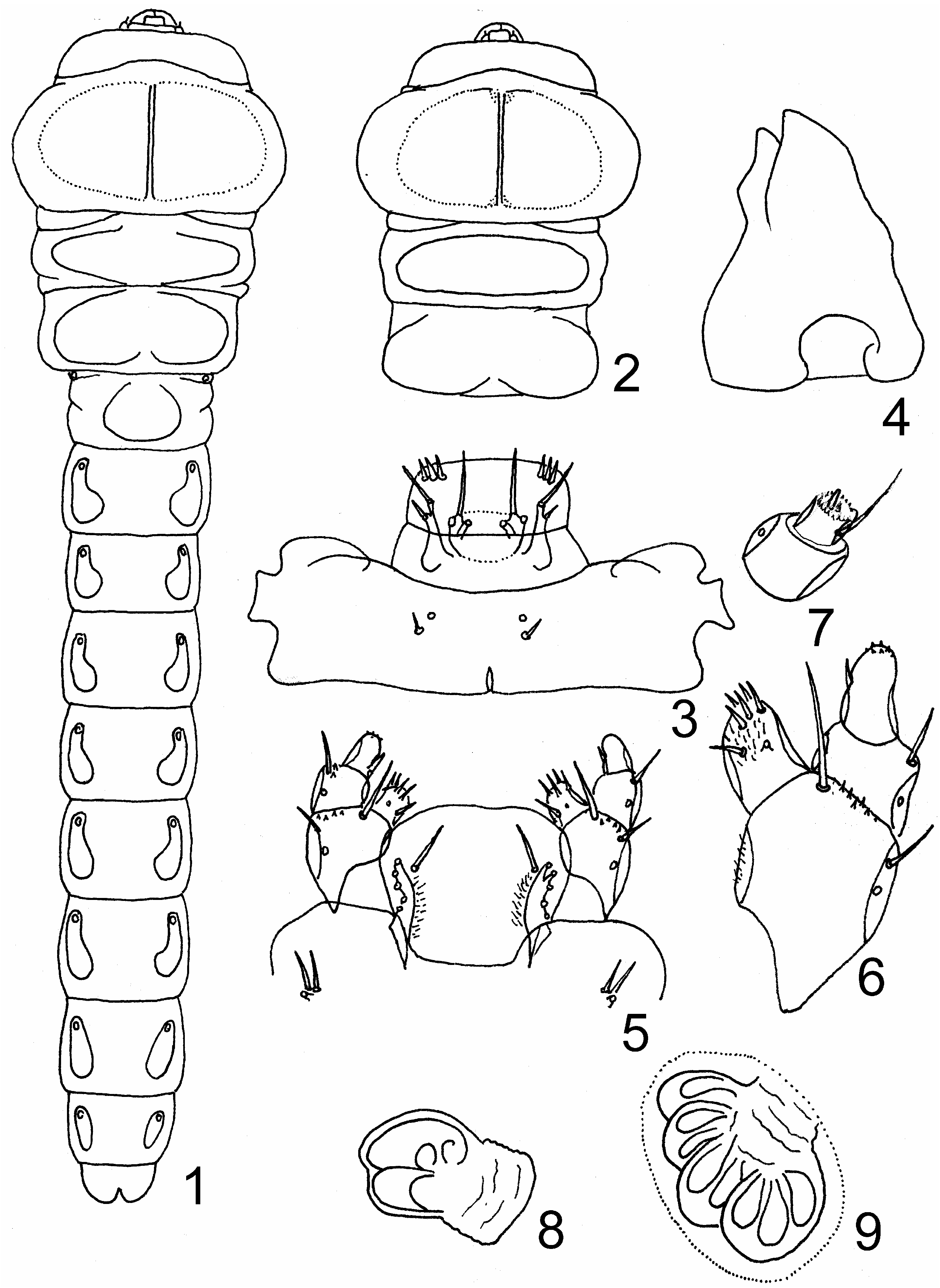
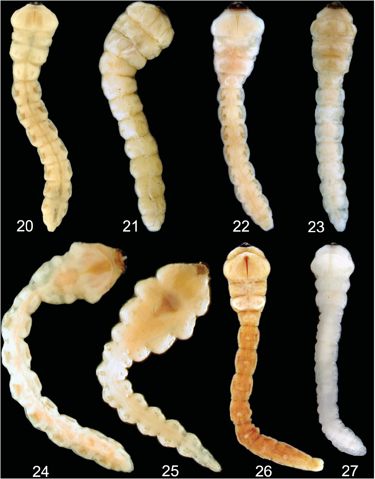
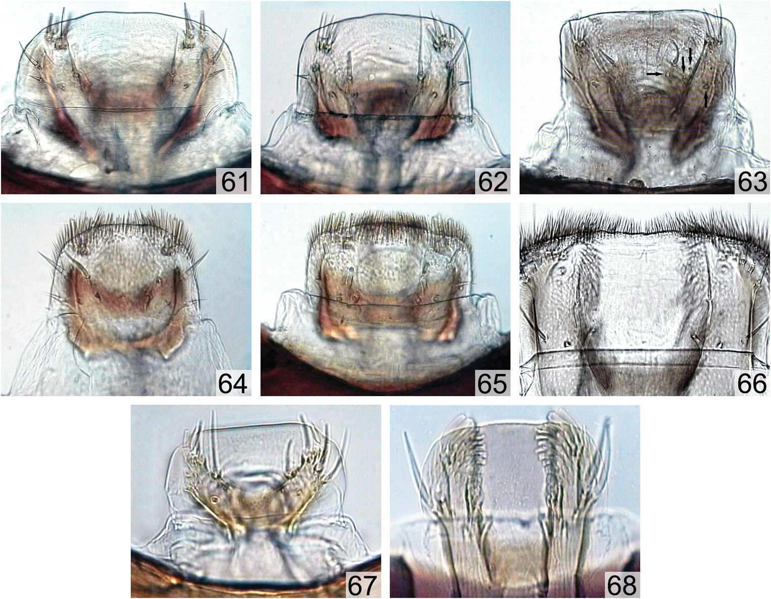
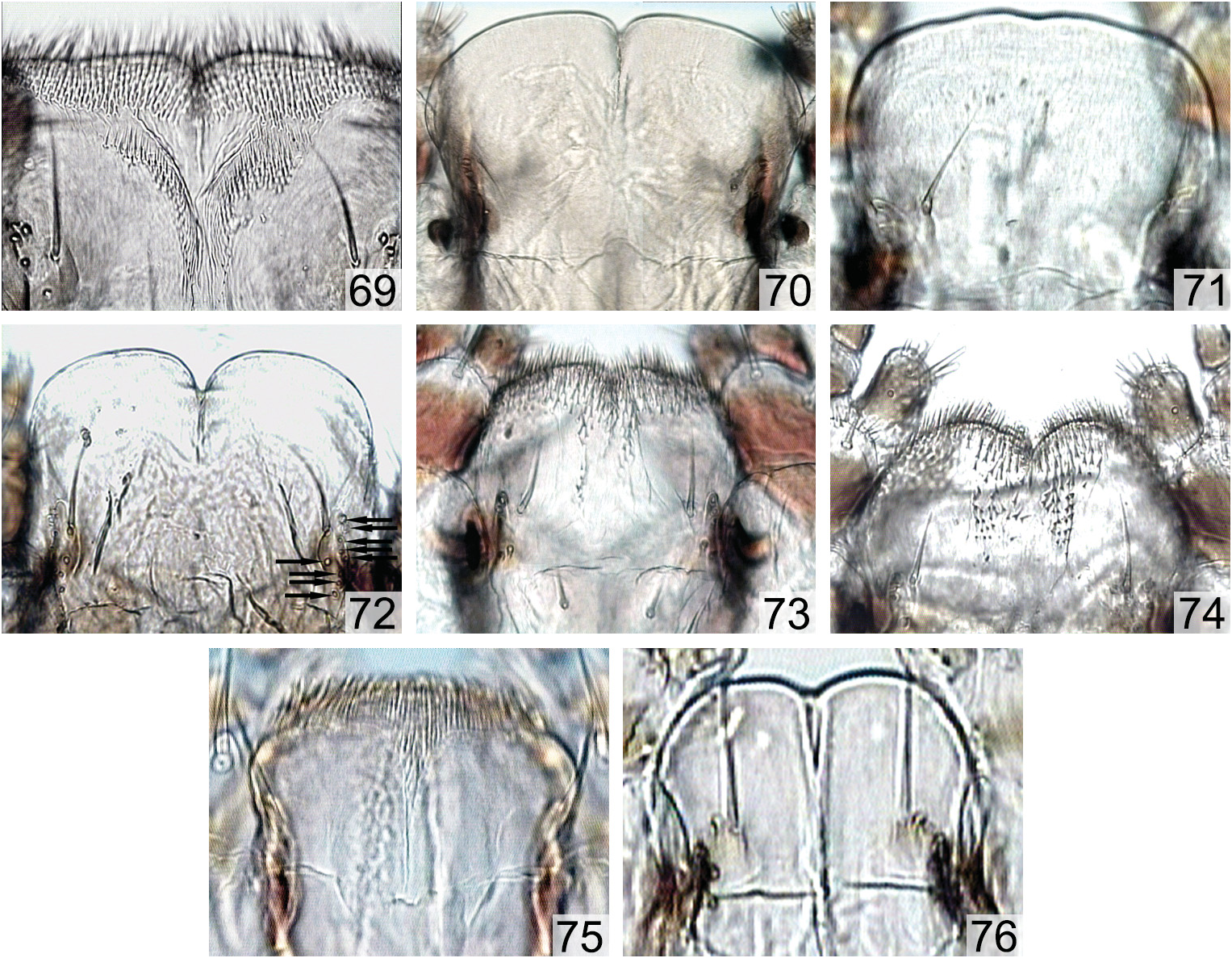
( Figs 1–9 View Figs 1–9 , 20 View Figs 20–27 , 28–29 View Figs 28–43 , 44, 53 View Figs 44–60 , 61 View Figs 61–68 , 70 View Figs 69–76 , 77, 85, 114–115)
Material examined. AUSTRALIA: WESTERN AUSTRALIA: Peak Chartes N.P., 8.xi.2001, S. Bílý leg., Eucalyptus sp. (Myrtaceae) , at the base of burned tree, 3 mature specimens ( 2 in NMPC, 1 in ZIN).
Description. Measurements ( 1 larva): body length 30.9 mm; width of prothorax 6.2 mm.
Larva of buprestoid type ( Figs 1 View Figs 1–9 , 20 View Figs 20–27 ), morpho-ecological subtype 2 ( VOLKOVITSH 1979, BÍLÝ 1999), prothorax poorly expanded, slightly wider than meso- and metathorax; body creamy-white, sides of thorax with sparse, short whitish setae, sides of abdomen with inconspicuous setae.
Head. Epistome ( Fig. 3 View Figs 1–9 ) strongly transverse, 5.9 times as wide as long; anterior margin deeply emarginate between oval mandibular condyles, with distinct rounded projections laterally; posterior margin bisinuous, distinctly emarginate laterally; latero-posterior corners nearly rectangular, projecting outwards; lateral margins of epistome slightly emarginate; antennal incisions broad, well-defined; 4 epistomal sensilla arranged in two superficial groups of two sensilla situated nearly linearly at midlength, each group consists of one short seta and one campaniform sensillum just above seta; distance between sensilla in each group much less than that between the groups. Clypeus ( Figs 3 View Figs 1–9 , 61 View Figs 61–68 ) strongly transverse, 3.2 times as wide as long, membranous, glabrous.
Labrum ( Figs 3 View Figs 1–9 , 61 View Figs 61–68 ) slightly transverse, 1.2 times as wide as long, anterior margin feebly bisinuous, middle part nearly straight, lateral lobes not developed; lateral sides of labrum widely arcuate, slightly emarginate at base, subparallel; palatine sclerites with well-defined separated branches; medial branches transverse, moderately sclerotised; lateral branches well-sclerotised, strongly expanded at bases; medial sensilla of labrum (t = trichoid, c = campaniform): 1c-2t-3c, distance 1c–3c more than 1c– 2t; 2t relatively short, not reaching anterior margin; antero-lateral sensilla of labrum, external group: ( 1t, 2c)- 3t + 4t; 1t and 3t relatively short but much longer than 4t, internal group: 1c-( 2t, 3t, 4t); labrum dorsally glabrous, ventrally (epipharynx) with large oblique areas of microspinulae along branches of palatine sclerite.
Antennae ( Fig. 7 View Figs 1–9 ) two-segmented, situated in very deep incision between epistome and pleurostome, basal antennomere 1.2 times as long as terminal antennomere; articular membrane glabrous; basal antennomere nearly as long as wide, barrel-shaped, with glabrous anterior margin; terminal antennomere slightly elongate, 1.3 times as long as wide at base, feebly expanded anteriorly, anterior margin with sparse microspinulae; apical cavity of terminal antennomere very shallow, bearing long trichosensilla, nearly 1.5 times as long as antennomere itself, arising from small separate cavity on external wall; sensory appendage elongate, conical, extending beyond anterior margin of cavity; two very short palmate sensilla with jointed bases and two basiconic sensilla of different length.
Mandibles ( Figs 4 View Figs 1–9 , 53 View Figs 44–60 ) triangular, 1.2 times as long as wide, strongly sclerotised at apical half, black, basal half lighter, brown; outer margin with one short seta above condyle; cutting edge with five teeth: apical tooth rather sharp, two internal teeth dorsally and ventrally both arising from common base.
Hypostome. Internal margin of mandibular fossae forms a big tooth-like projection, nearly reaching latero-basal sclerite of cardo ( Fig. 44 View Figs 44–60 ).
Maxillae ( Figs 5, 6 View Figs 1–9 , 77, 85). Cardo subquadrate, nearly as wide as long, membranous, glabrous; latero-basal sclerite of cardo (Fig. 77) well-defined and sclerotised, bearing two short setae and one campaniform sensillum with enlarged bases. Stipes ( Figs 6 View Figs 1–9 , 85) subquadrate, slightly wider than long; inner sclerite of stipes well-defined, with internal process very short; apical seta of stipes slightly longer than basal palpomere, situated at anterior 1/3 of stipes length between the bases of palpus and mala; lateral seta very short, situated at apex of external part of inner sclerite; external armament composed of small area of sparse, short microspinulae at antero-external corners, anterior margin glabrous, internal armament composed of sparse, short microspinulae laterally, extending mala; additional internal lobe absent. Palpus maxillaris ( Figs 6 View Figs 1–9 , 85): basal palpomere nearly as long as terminal palpomere; basal palpomere trapezoid, slightly transverse, 1.1 times wider than long, completely glabrous, apical seta nearly twice shorter than terminal palpomere; terminal palpomere subcylindrical, weakly narrowed and curved outward apically, 1.4 times as long as wide, with approximately ten short, peg-like apical sensilla, one of them larger than others. Mala ( Figs 6 View Figs 1–9 , 85) slightly elongate, feebly narrowed apically, 1.2 times as long as wide; inner sclerite of mala well-defined and sclerotised, subparallel, slightly longer than wide; external armament of mala composed of one campaniform sensillum, 1 short, peg-like seta, five long robust setae, and short microspinulae apically; mala internally with 3 robust, short setae and dense microspinulae along inner side.
Labium ( Figs 5 View Figs 1–9 , 70 View Figs 69–76 ). Prementum transverse, 1.6 times as wide as long, rounded; anterior margin widely arcuate, deeply emarginate at middle, with broadly rounded corners; lateral sides widely arcuate, strongly converging posteriorly; prementum completely glabrous both dorsally and ventrally; corner sclerites of labium with poorly defined anterior part, apical seta relatively long but not extending anterior margin of prementum; five campaniform sensilla situated below the base of apical setae, three apical (two with long bases) and two basal. Postmentum glabrous, without setae.
Thorax ( Figs 1–2 View Figs 1–9 , 20 View Figs 20–27 , 28–29 View Figs 28–43 ). Slightly expanded, flattened, prothorax 1.2 times wider than mesothorax which is nearly as wide as metathorax and both slightly wider than 1 st abdominal segment; rudiments of legs present ventrally on all thoracic segments. Prothorax transverse, 1.8 times as wide as long; anterior membrane glabrous with sparse short setae and inconspicuous microspinulae medially; lateral sides evenly rounded, with moderately dense, short, whitish setae. Both dorsal and ventral plates poorly defined, unsclerotised, colorless, limited by oblique folds. Pronotal plate ( Figs 1 View Figs 1–9 , 28 View Figs 28–43 ) glabrous, bearing sparse short setae and grainy superficial cuticular sculpture along the groove. Pronotal groove uniramous, widest at apex, gradually narrowed toward base where it is forked; lateral branches short, curved outwards. Prosternal plate ( Figs 2 View Figs 1–9 , 29 View Figs 28–43 ) glabrous, bearing sparse short setae, grainy superficial cuticular sculpture along the groove, and a few concentric rugae. Prosternal groove uniramous, inverted T-shaped, widest at apex, becoming narrower toward base where it shortly triangularly widened forming short, nearly perpendicular branches. Mesothorax ( Figs 1–2 View Figs 1–9 , 28–29 View Figs 28–43 ) transverse, 2.9 times as wide as long and 1.1 times wider than metathorax, with well-defined secondary fold dividing segment into two parts; glabrous, bearing sparse short setae, with hardly visible areas of poorly developed microspinulae laterally; ambulatory pads well-defined dorsally and ventrally, ovale. Metathorax ( Figs 1, 2 View Figs 1–9 , 28, 29 View Figs 28–43 ) transverse, 2.2 times as wide as long and 1.4 times wider than 1 st abdominal segment; lateral sides with sparse short whitish setae; both dorsal and ventral sides of metathorax with distinct, transverse plates covered with microspinulae; dorsally with two large ambulatory pads separated by median depression and bearing inconspicuous cuticular sculpture laterally, ventrally with single, large, transverse, reniform medial pad.
Abdomen ( Figs 1 View Figs 1–9 , 20 View Figs 20–27 ) flattened, segments 1–8 weakly transverse, slightly wider than long; all segments laterally, segments 4–10 dorsally and 6–10 ventrally with microspinulate areas; lateral sides with sparse short whitish setae and poorly defined microspinulae; 1 st abdominal segment trapezoid, narrowed posteriorly, 1.4 times as wide as long and narrower than 2 nd segment; dorsally with large ambulatory pad divided by shallow longitudinal depression, bearing rugulose sculpture; ventrally with large, medial ambulatory pad divided by transverse furrow into two parts, ends of furrow with inconspicuous inner sculpture; mainly glabrous, with sparse short setae; sides covered with microspinulae. Abdominal segments 2–8 slightly transverse, 1.3–1.6 times as wide as long; 9 th abdominal segment slightly transverse, arcuately narrowed posteriorly, 1.4 times as wide as long; 10 th abdominal segment conical, 1.4 times as wide as long, with vertical unsclerotised anal rim.
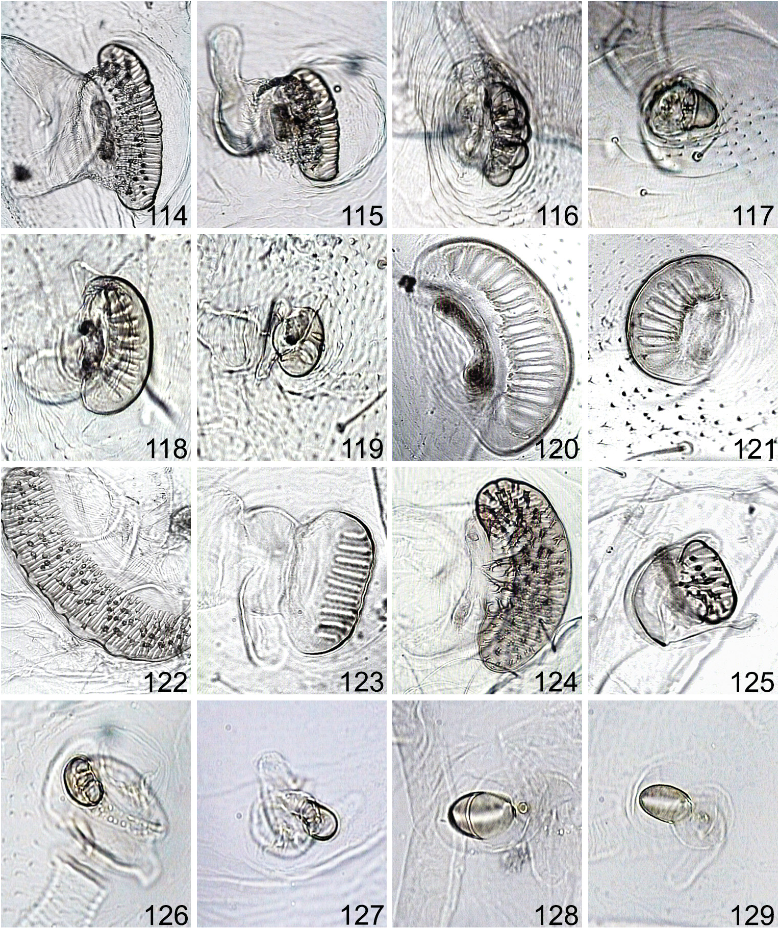
Spiracles ( Figs 8–9 View Figs 1–9 , 114–115 View Figs 114–129 ). Mesothoracic spiracles ( Figs 9 View Figs 1–9 , 114 View Figs 114–129 ) of buprestoid, cribriform type, reniform, narrow, strongly transverse, 3.3 times as wide as long, situated on sides of anterior part of mesothorax, with cancellate peritreme and numerous, branched trabeculae; spiracles surrounded with poorly defined microspinulae. Abdominal spiracles ( Figs 8 View Figs 1–9 , 115 View Figs 114–129 ) of the same type, renifom, narrow, transverse (that on 1 st abdominal segment 2.7 times as wide as long), situated dorsally in depressions on abdominal segments 1–8, with cancellate peritreme and numerous branched trabeculae; adjacent cuticle glabrous or spiracles surrounded with poorly defined microspinulae.
Proventriculus (Figs 106–107) with complicate inner armament consisting of different elements and moderately developed dorsal and ventral central stripes; nearly globular, with two long, curved gastric caeca at base which are about 1.5 times longer than proventriculus itself; main fields with very dense, robust, singular microteeth situated on tubercles (Fig. 106); their margins with smaller and sparser, reduced teeth and enlarged tubercles, anteriorly with small fine microspinulae situated in rows, posteriorly with very long microspinulae changing to microteeth. Central stripes developed on both sides; dorsally incomplete, not extending apex, at base with long and dense microspinulae (Fig. 107) changing to shorter microspinulae with expanded basal tubercles then, at the middle, to short sparse microteeth, and, anteriorly, to long slender and dense fine setae; ventrally with short posterior area limited with narrow glabrous stripes, bearing long and short microspinulae directed medially and posteriorly and surrounding the field of short inconspicuous microspinulae situated in 2–3 on common bases. Glabrous areas extensive, with long wavy rugosities.


No known copyright restrictions apply. See Agosti, D., Egloff, W., 2009. Taxonomic information exchange and copyright: the Plazi approach. BMC Research Notes 2009, 2:53 for further explanation.