
Watersipora subovoidea (d’Orbigny, 1852), d'Orbigny, 1852
|
publication ID |
https://doi.org/10.5281/zenodo.274831 |
|
DOI |
https://doi.org/10.5281/zenodo.3507454 |
|
persistent identifier |
https://treatment.plazi.org/id/03E6A433-576D-FFA6-6E9D-1364FE62FE9A |
|
treatment provided by |
Plazi (2016-04-19 13:33:32, last updated 2024-11-28 08:27:52) |
|
scientific name |
Watersipora subovoidea (d’Orbigny, 1852) |
| status |
|
Watersipora subovoidea (d’Orbigny, 1852) View in CoL
( Fig. 4 View FIGURE 4 C, D, G, H)
Cellepora subovoidea d’Orbigny, 1852: 402 .
Lepralia cucullata Busk, 1854: 81 , pl. 96, figs 4, 5.
Schizoporella cucullata View in CoL var. labiosa Calvet, 1903: 141 –142, pl. 16, fig. 7a–c.
Smittia (Watersipora) cucullata: Neviani 1895: 83 .
Lepralia View in CoL ? cucullata: Waters 1909: 150 , pl. 15, figs 2–4 [pars].
Watersipora cucullata: Hastings 1930: 729 View in CoL [pars], pl. 15, figs 97–101 (not 102–104); Soule & Soule 1976: 306, pl. 2, figs 1–2; pl. 3, fig. 1; pl. 4, fig. 3; also pl. 3, fig. 2, as Cellepora ovoidea , from Savigny’s (1817) plate.
[? Not] Dakaria subovoidea: Harmer 1957: 1022 , pl. 69, figs 11, 12, 14.
Watersipora subovoidea: Gautier 1962: 183 View in CoL ; Ryland 1965: 68, figs 33c, d; 1974: 345, fig. 3A; Soule & Soule 1976: 302, pl. 3, fig. 4; pl. 4, fig. 4; Zabala 1986: 396, fig. 129; Hayward & McKinney 2002: 63, fig. 29A, B.
Material examined. All specimens are in the Natural History Museum, London. Naples, Italy, 20-10-1960, JSR (1 slide, intact, NHM 2007.12.14.2); Arrawarra, New South Wales, Australia, January 1772, JSR (3 slides: 1 cleaned, NHM 2007.12.14.3; 2 decalcified, NHM 2007.12.14.4–5) ( Ryland 1974); Port of Genoa, Italy, 12-11-2007, M. Faimali (3 slides: 1 cleaned, NHM 2007.12.14.6; 2 decalcified, NHM 2007.12.14.7–8).
Description. Colonies encrusting, unilaminar, multilaminar or sometimes foliaceous, brownish-purple to almost black in life (fading to grey when dried), with the growing margin orange. Zooids subrectangular or slightly hexagonal, sometimes narrower proximally (usually when associated with row division); large, distinct, about twice as long as wide, 700–1100 (mean ~900) × 350–650 (mean ~450) μm (globally; Table 2 View TABLE 2 ), variable within and between colonies. Frontal skeletal wall rather flat, perforated by numerous large round pseudopores 20–30 μm diameter; in some zooids two or more distal pores, largely concealed by the orificial rim, may be areolar septular pores; covered by a variably pigmented layer and a shiny, transparent cuticle. Orifice large, subcircular to oval, slightly wider than long; 200–250 (mean ~205) × 190–250 (mean ~230) μm; occupying <10% of the total zooid area; with a proximal sinus demarcated by the condyles, ~50 × 100 μm; surrounding rim variably elevated. Condyles triangular, projecting tooth-like distomedially into the anter (distal portion of orifice). Operculum strongly pigmented, with a broad, parallel-sided dark central band demarcated by sclerites, paler peripheral regions, and two white spots (lucidae) proximally, each adjacent to the condyle. 23–24 tentacles ( Waters 1909, Naples).
Variation and remarks. The rim of the orifice may be somewhat elevated on either side of the sinus, as in the form distinguished as var. labiosa by Calvet (1903, pp. 141, 142), from the intertidal of Graciosa, Azores. Waters (1909) confounded two species under his Lepralia cucullata ― his Naples specimens were correctly identified and are now therefore referred to W. subovoidea , but those from the Red Sea (Plate 15, figs 1, 5), later distinguished as var. watersi by Mawatari (1952), are clearly referable on the basis of the operculum to W. subtorquata .
Calvet, L. (1903) Deuxieme Partie. In: Jullien, J. & Calvet, L. Bryozoaires provenant des campagnes de l' Hirondelle (1886 - 1888). Resultats des campagnes scientifiques accomplies sur son yacht par Albert 1 er, Prince Souverain de Monaco, 1903, 23, 1 - 188.
Gautier, Y. V. (1962) Recherches ecologiques sur les Bryozoaires chilostomes en Mediterranee occidentale. Receuil des Travaux de la Station marine d'Endoume, Fasc. 38, Bull. 24, 1 - 434.
Harmer, S. F. (1957) The Polyzoa of the Siboga Expedition. Part 4. Cheilostomata Ascophora II. Siboga Expeditie, 28 D, 641 - 1147.
Hastings, A. B. (1930) Cheilostomatous Polyzoa from the vicinity of the Panama Canal collected by Dr. C. Crossland on the cruise of the S. Y. ' St. George'. Proceedings of the Zoological Society of London, 1929, 697 - 740.
Hayward, P. J. & McKinney, F. K. (2002) Northern Adriatic Bryozoa from the vicinity of Rovinj, Croatia. Bulletin of the American Museum of Natural History, 270, 1 - 139.
Mawatari, S. (1952) On Watersipora cucullata (Busk). I. Systematic study. Miscellaneous Reports of the Research Institute of Natural Resources (Tokyo), 25, 14 - 17.
Neviani, A. (1895) Briozoi fossili della Farnesina e Monte Mario presso Roma. Palaeontographia italica, 1, 77 - 140.
Ryland, J. S. (1965) Polyzoa. OECD Catalogue of Main Marine Fouling Organisms, 2, 1 - 83.
Ryland, J. S. (1974) Bryozoa in the Great Barrier Reef province. In: Proceedings of the Second International Reef Symposium Volume 2. Cameron, A. M. et al. Brisbane, The Great Barrier Reef Committee. Pp. 341 - 348.
Soule, D. F. & Soule, J. D. (1976) Species groups in Watersiporidae. Bryozoa 1974. Documents des Laboratoires de Geologie de la Faculte des Science de Lyon, h. s. 3, 2, 299 - 309.
Waters, A. W. (1909) Cheilostomata. Reports on the marine biology of the Sudanese Red Sea. XII. The Bryozoa. Part I. Cheilostomata. Journal of the Linnean Society (Zoology), 31, 123 - 181.
Zabala, M. (1986). Fauna dels Briozous dels Paisos Catalans. Barcelona, Institut d'Estudis Catalans. 833 pp.
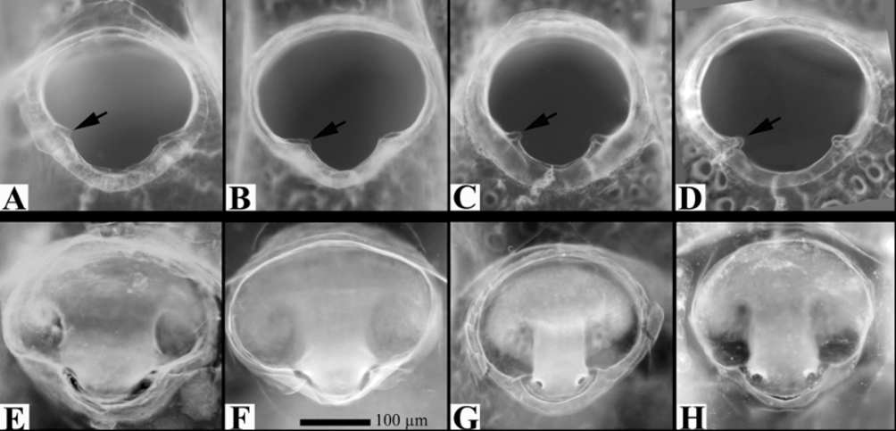
FIGURE 4. Light photomicrographs of orifices and opercula of Watersipora. Scale bar = 100 Μm. A – D, orifices; E – H, opercula. A. W. subtorquata, St-Jacut-de-la-Mer, France; B. W. subtorquata, St Peter Port, Guernsey; C. W. subovoidea, Arrawarra, New South Wales, Australia; D. W. subovoidea, Genoa, Italy; E. W. subtorquata, St-Jacut-de-la-Mer, France; F. W. subtorquata, St Peter Port, Guernsey; G. W. subovoidea, Naples, Italy; H. W. subovoidea, Genoa, Italy. In A – D, note the differences in condyles (the left one in each picture arrowed); in E – H, compare the sides of the central dark band, concave in W. subtorquata, parallel in W. subovoidea.
No known copyright restrictions apply. See Agosti, D., Egloff, W., 2009. Taxonomic information exchange and copyright: the Plazi approach. BMC Research Notes 2009, 2:53 for further explanation.
|
Kingdom |
|
|
Phylum |
|
|
Class |
|
|
Order |
|
|
Family |
|
|
Genus |
Watersipora subovoidea (d’Orbigny, 1852)
| Ryland, John S., Blauwe, Hans De, Lord, Richard & Mackie, Joshua A. 2009 |
Watersipora subovoidea:
| Hayward 2002: 63 |
| Zabala 1986: 396 |
| Soule 1976: 302 |
| Ryland 1965: 68 |
| Gautier 1962: 183 |
Watersipora cucullata:
| Soule 1976: 306 |
| Hastings 1930: 729 |
Lepralia
| Waters 1909: 150 |
Schizoporella cucullata
| Calvet 1903: 141 |
Smittia (Watersipora) cucullata:
| Neviani 1895: 83 |
1 (by plazi, 2016-04-19 13:33:32)
2 (by ImsDioSync, 2017-02-08 14:33:28)
3 (by ExternalLinkService, 2019-09-26 12:12:25)
4 (by ExternalLinkService, 2019-10-18 14:46:37)
5 (by ExternalLinkService, 2021-11-09 19:33:11)
6 (by ExternalLinkService, 2021-11-10 11:37:54)
7 (by plazi, 2023-10-29 13:12:02)