
Drosophila neocordata Magalhães, 1956
|
publication ID |
https://doi.org/10.11646/zootaxa.5061.3.7 |
|
publication LSID |
lsid:zoobank.org:pub:8C2F06C6-BF5C-450F-8098-66CEE68709BC |
|
DOI |
https://doi.org/10.5281/zenodo.5649956 |
|
persistent identifier |
https://treatment.plazi.org/id/03E26406-0239-4663-839A-B49280FEFE2B |
|
treatment provided by |
Plazi |
|
scientific name |
Drosophila neocordata Magalhães, 1956 |
| status |
|
Drosophila neocordata Magalhães, 1956 View in CoL
( Fig. 16 View FIGURE 16 )
Non-type material. Strain CG ( Campo Grande , Mato Grosso do Sul, Brazil): 12 males dissected .
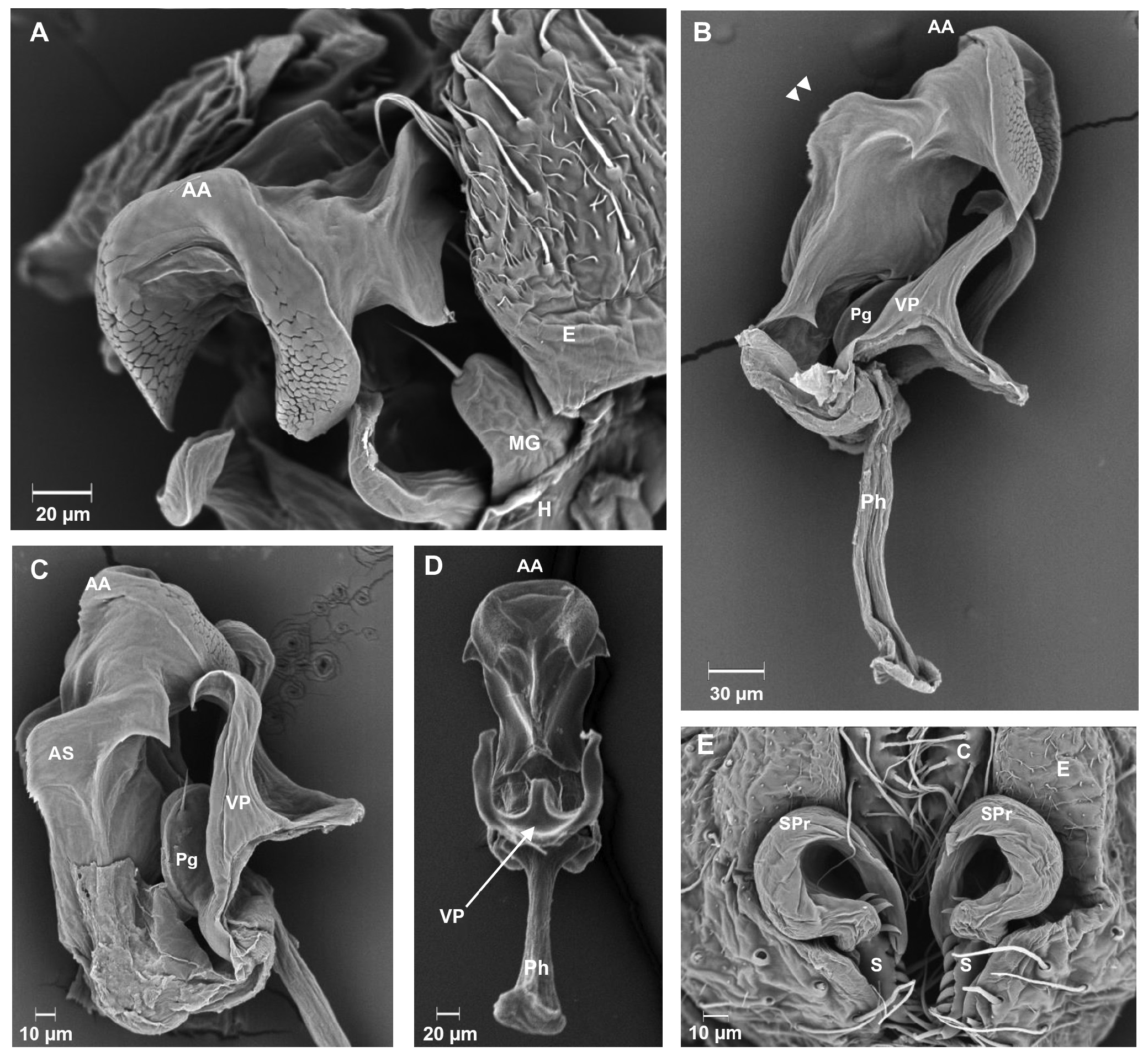
Male terminalia. The dorso-ventral region of the epandrium is angular. The epandrium is covered with short and long epandrial bristles ( Fig. 16E View FIGURE 16 ). The cercus is U-shaped, covered with cercal bristles ( Fig. 16E View FIGURE 16 ). The surstyli is concave and has a row of 5 to 6 surstylar teeth arranged throughout the internal portion of each surstylus and a tuft of long surstylar bristles at the inferior region ( Fig. 16E View FIGURE 16 ). Each surstylus of this species have a unique characteristic, not seen in other subgroups, the surstylar process. This structure is found connected to each surtylus and it has surstylar teeth at its ends ( Fig. 16E View FIGURE 16 ). The hypandrium is small and it has a median gonocoxites, with the presence of a hypandrial bristle at each end ( Fig. 16A View FIGURE 16 ). The aedeagus presents a pair of chitinous hooks in the frontal region of the aedeagal apex, which extend to the ventral region of the aedeagus body ( Fig. 16A–D View FIGURE 16 ). There is a pair of long protuberances, serrated at the edge, arranged laterally, and fused to the aedeagus; we suggest that these protuberances can be the aedeagal sheath partly fused to the aedeagus body ( Fig. 16B, C View FIGURE 16 ). The pregonites are more different, they seem to be fused into a single structure, and present two small pregonal bristles ( Fig. 16B, C View FIGURE 16 ). The ventral postgonites are long, thin, and bifurcated in the middle region; they are also folded inward at their ends, which fit into the chitinous hooks ( Fig. 16B–D View FIGURE 16 ). The pregonite and the ventral postgonites are connected to the phallapodeme ( Fig. 16B–D View FIGURE 16 ). The phallapodeme is short and thin ( Fig. 16B, D View FIGURE 16 ).
| CG |
Embrapa Collection of Fungi of Invertebrates |
No known copyright restrictions apply. See Agosti, D., Egloff, W., 2009. Taxonomic information exchange and copyright: the Plazi approach. BMC Research Notes 2009, 2:53 for further explanation.
|
Kingdom |
|
|
Phylum |
|
|
Class |
|
|
Order |
|
|
Family |
|
|
Genus |