
Armillipora Quate, 1996
|
publication ID |
https://doi.org/10.11646/zootaxa.4890.3.8 |
|
publication LSID |
lsid:zoobank.org:pub:E9E1172F-59CB-48DF-927A-261D3C324CCB |
|
DOI |
https://doi.org/10.5281/zenodo.4328523 |
|
persistent identifier |
https://treatment.plazi.org/id/03D68795-FFB6-FFB6-92F1-95D9FADDFE77 |
|
treatment provided by |
Plazi |
|
scientific name |
Armillipora Quate |
| status |
|
Genus Armillipora Quate View in CoL
Armillipora Quate, 1996: 29 View in CoL . Type species: Armillipora selvica Quate, 1996 View in CoL (by orig. des.)
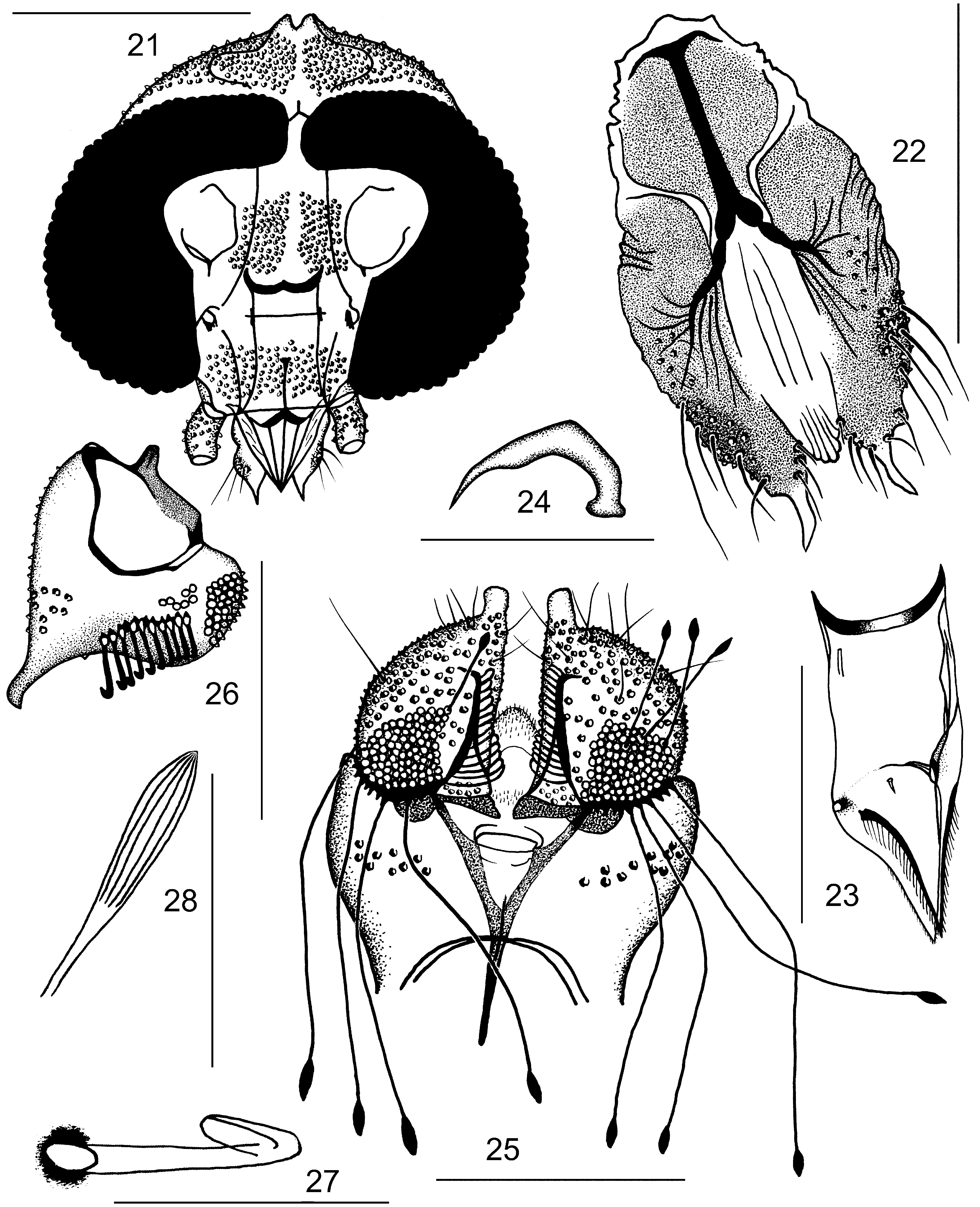
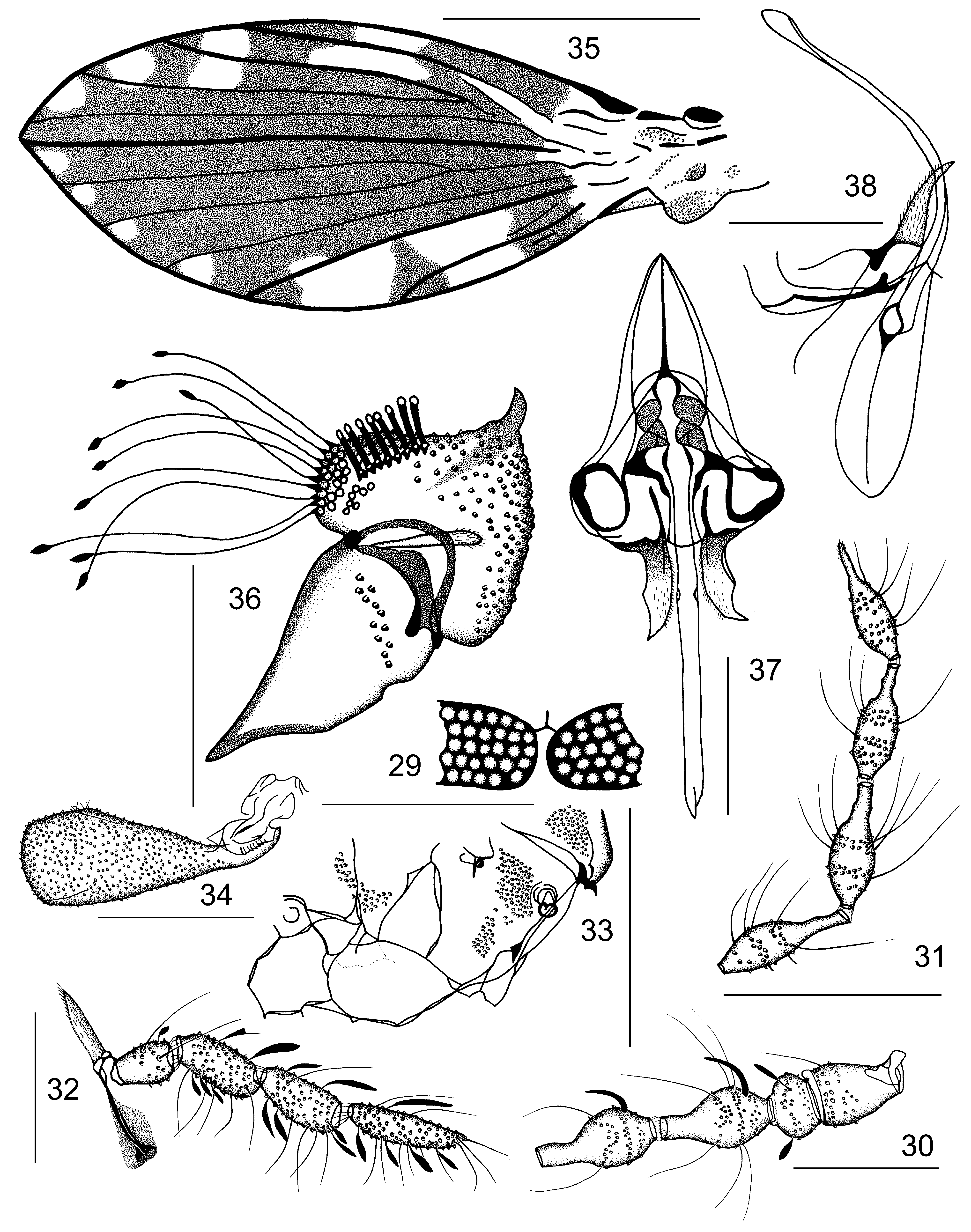
Redescription of males. Armillipora is characterized by the following characters: Head ( Figs. 1 View Figures 1–10 , 21 View Figures 21–28 ) in horizontal axis a little broader than in vertical axis, flattened antero-posteriorly, vertex rounded, occipital lobulus with an indentation apically. Corniculi not developed; insertions of supraocular bristles on dorsal margins of eyes not enlarged. Scars on vertex divided by a median, scar free band. Eyes separated, eye bridge formed by 4–5 facet rows, the number of facets in the apices being more reduced. The eye bridge is narrower than the ventral portion of the eye. Interocular frontal suture sclerotized ( Figs. 1 View Figures 1–10 , 11 View Figures 11–20 , 21 View Figures 21–28 , 29 View Figures 29–38 ), Y-shaped. Frontoclypeus ( Figs. 1 View Figures 1–10 , 21 View Figures 21–28 ) with two vertical, quite separated, approximately oblong, alveoli patches. Antennae ( Figs. 12 View Figures 11–20 , 30, 31 View Figures 29–38 ) with scape truncated, narrower at the base and widened distally, pedicel globular with scales very narrow and of different lengths. Flagellomeres pitcher-shaped, symmetrical, with necks shorter than swollen basal parts, with pores around each node, apiculus of the last flagellomere developed ( Fig. 31 View Figures 29–38 ). Sensory filaments (ascoids) simple, paired and needle-shaped ( Figs. 12 View Figures 11–20 , 30 View Figures 29–38 ). Last segment of maxillary palpus not annulated ( Figs. 2 View Figures 1–10 , 32 View Figures 29–38 ). Terminal lobes of labium with apical pointed projection ( Figs. 3 View Figures 1–10 , 22 View Figures 21–28 ).
Thorax. Anepisternum setae patch composed of two quite separated groups (not mentioned by Quate), anepimeron setae patch divided, but not always so markedly ( Fig. 5 View Figures 1–10 , 33 View Figures 29–38 ). Metathoracic spiracles set low on mesothorax. There are no thoracic allurement organs. Wings ( Figs. 13 View Figures 11–20 , 35 View Figures 29–38 ) broadly lanceolate, pointed at the end of R 5, somewhat expanded at the posterior margin and characteristically maculated; three-quarters of wing membrane dark. Radial and medial fork complete, basal to the wing centre, positioned as seen in Figs. 13 View Figures 11–20 , 35 View Figures 29–38 . Medial wing angle 167–180°.
Male terminalia. Aedeagal complex symmetrical. Gonocoxal condyles ( Figs. 17 View Figures 11–20 , 38 View Figures 29–38 ) penetrate a concavity on the underside of basiphallus. Epandrium almost square-shaped, setae are very sporadic on both sides of the central aperture ( Figs. 8 View Figures 1–10 , 14 View Figures 11–20 , 25 View Figures 21–28 , 36 View Figures 29–38 ). Hypandrium on both sides narrow and sclerotized, however, membranous in the middle. Remainders of ventral epandrial plate in the form of two gradually tapering strings to the sclerotized tip. Epandrial clasping lobes ( Figs. 8 View Figures 1–10 , 14 View Figures 11–20 , 25, 26 View Figures 21–28 , 36 View Figures 29–38 ) approximately hemispherical, haired, prolonged and tapering distally with a spoon-shaped hairless protuberance and dorsally provided by two tubercles with quite different shapes of tenacula: cylindrical ( Figs. 8 View Figures 1–10 , 14, 15 View Figures 11–20 , 25, 26, 27 View Figures 21–28 , 36 View Figures 29–38 ) with a folded terminal part and a very long accessory type with lancet-shaped tips ( Figs. 8 View Figures 1–10 , 14, 16 View Figures 11–20 , 25, 28 View Figures 21–28 , 36 View Figures 29–38 ).
Differential diagnosis. Probably a neotropical genus of Maruinini Enderlein, 1937 (compare Duckhouse 1987, 1990, Quate 1996, Ibáñez-Bernal & Suárez-Landa 2015), by the presence of a wing with radial fork basal to medial one, and both placed rather basally on wing; by the shape of the basiphallus (ejaculatory apodeme), which is broad, dorso-ventrally flattened; and the gonocoxal condyles ( Figs. 17 View Figures 11–20 , 38 View Figures 29–38 ) fitting into a concavity on the underside of the basiphallus. Armillipora resembles, upon first view of a collector in nature, species of Alepia Enderlein, 1937 , and Platyplastinx Enderlein, 1937 , by its conspicuous wing maculation and less distinctly visible characters, such as the shape of two types of tenacula (short cylindrical with a folded tip and long accessory filaments with a lancetshaped ending). It clearly differs from genera of the former Setomimini of Quate & Brown (2004) by no detection of declined gonopods as well as the presence of parameres, both sometimes quite reduced or missing, with only a conspicuous, very long aedeagal complex. The tunica is not developed (compare to known species of Alepia ).
Included taxa. Armillipora selvica Quate, 1996 , and A. suapiensis Ježek, Oboňa & Le Pont sp. nov.


No known copyright restrictions apply. See Agosti, D., Egloff, W., 2009. Taxonomic information exchange and copyright: the Plazi approach. BMC Research Notes 2009, 2:53 for further explanation.
|
Kingdom |
|
|
Phylum |
|
|
Class |
|
|
Order |
|
|
Family |
|
|
SubFamily |
Psychodinae |
|
Tribe |
Maruinini |
Armillipora Quate
| Ježek, Jan, Oboňa, Jozef, Pont, Ois Le, Maes, Jean-Michel & Martinez, Eddy 2020 |
Armillipora
| Quate, L. W. 1996: 29 |