
Braunus prionotus Barão & Garbelotto, 2016
|
publication ID |
https://doi.org/10.11646/zootaxa.4078.1.16 |
|
publication LSID |
lsid:zoobank.org:pub:0102DC85-29D8-4B7A-BD24-984C115189AA |
|
persistent identifier |
https://treatment.plazi.org/id/03BF87AB-1B22-FFC3-9AC4-9FD4379DFF3C |
|
treatment provided by |
ImsDioSync |
|
scientific name |
Braunus prionotus Barão & Garbelotto |
| status |
sp. nov. |
Braunus prionotus Barão & Garbelotto sp. nov.
( Figures 48–53 View FIGURES 48 – 53 )
Material examined. Holotype ♀, COLOMBIA, VII. 1974, Steinheil leg. ( MNHN).
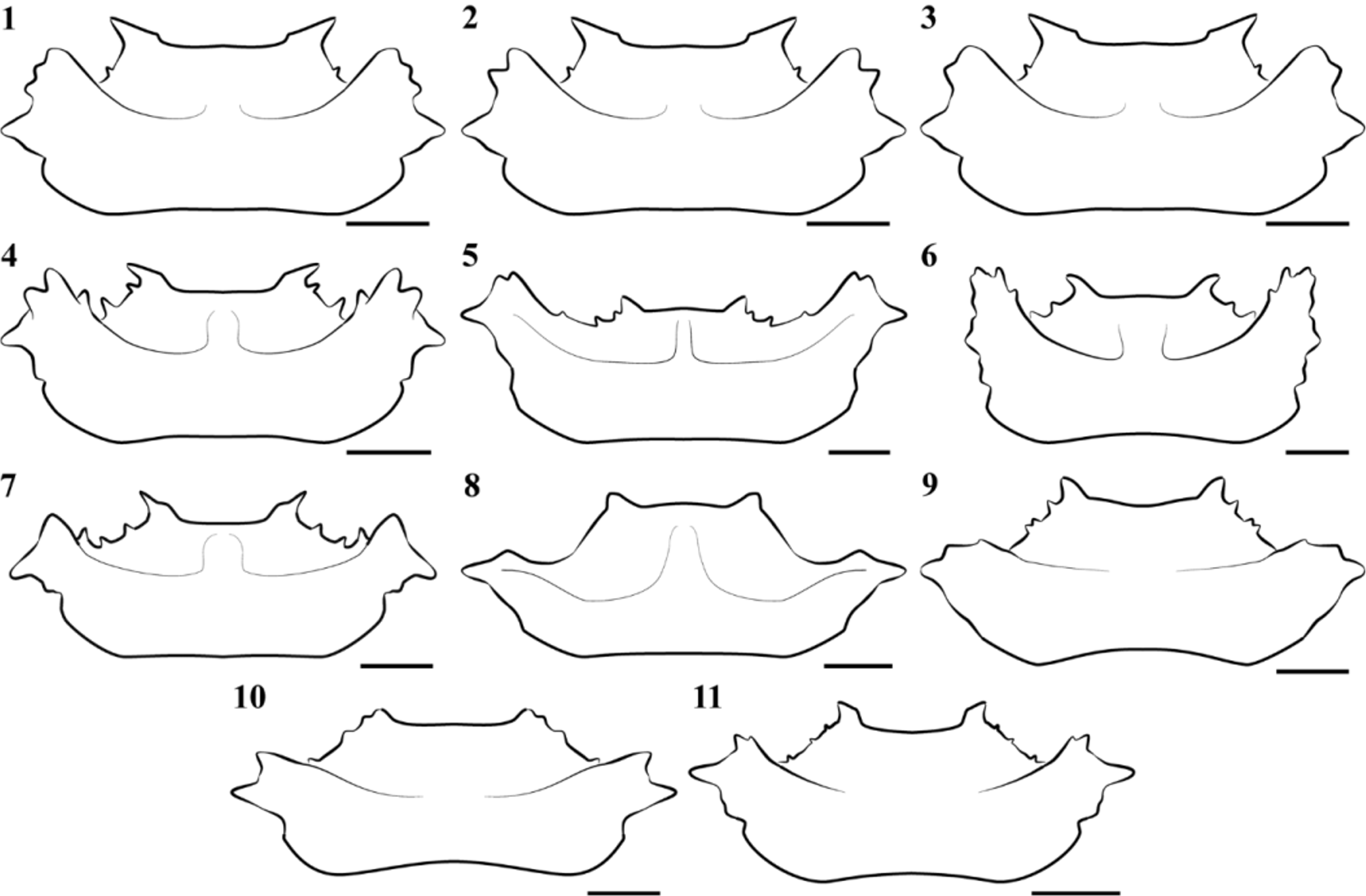
Diagnosis. Lateral margins of mandibular plates denticulate ( Fig. 48, 50 View FIGURES 48 – 53 ). Anterior angle of pronotum with an obtuse process; anterolateral margins with two small tubercles; humeral angles directed anteriad, each with multiple tubercles ( Fig. 6 View FIGURES 1 – 11 , 48 View FIGURES 48 – 53 ). Apex of hemelytral radial vein calloused; corium reaching abdominal segment VI. Apex of scutellum reflexed laterally. Female genitalia: gonocoxites 8 wider than long; posterior margin of each gonocoxite 8 concave; posterior margin of each laterotergite 8 obtuse; posterior margin of gonocoxites 9 concave ( Figs 52–53 View FIGURES 48 – 53 ).
Description. Body color dark brown ( Figs 48–49 View FIGURES 48 – 53 ). Mandibular plates longer than clypeus; lateral margins denticulate ( Fig. 48 View FIGURES 48 – 53 ). Proportion of antennal segments: I<II=IV<III (antennal segment V missing). Labial apex attaining abdominal sternite IV ( Fig. 49 View FIGURES 48 – 53 ). Processes at anterior angles of pronotum each directed laterally, obtuse apically, not reaching posterior margins of compound eyes. Anterolateral margins of pronotum each bearing 2 + 2 little-produced, finger-like tubercles; humeral angles projected anteriad, each with multiple processes; each posterolateral margin sinuate; pronotal disc calloused between cicatrices ( Fig. 6 View FIGURES 1 – 11 ). Scutellum reaching posterior margin of abdominal segment VI; scutellum swollen longitudinally, medially; postfrenal lobe about 1 / 2 of length of frenal lobe; apex of scutellum reflexed laterally. Hemelytral corium reaching abdominal segment VI; radial vein calloused apically; membrane reduced, not surpassing abdominal segment VI, veins absent. Hind wings reduced.
Female genitalia. Gonocoxites 8 wider than long; each sutural margin parallel and juxtaposed at 1 / 3 basal portion, widely separated and concave on the remaining 2 / 3; posterior margins concave ( Figs 52–53 View FIGURES 48 – 53 ; gc 8). Gonapophyses 8 visible, subtriangular, with median carina ( Figs 52–53 View FIGURES 48 – 53 ; g 8). Laterotergites 8 triangular, longer than wide, each posterior margin obtuse, surpassing tergite 8 ( Figs 52–53 View FIGURES 48 – 53 ; la 8). Gonocoxites 9 trapezoidal, posterior margin convex ( Figs 52–53 View FIGURES 48 – 53 ; gc 9). Laterotergites 9 finger-like, each strongly depressed basally; apices rounded, each barely surpassing posterior margin of tergite 8 ( Figs 52–53 View FIGURES 48 – 53 ; la 9). Segment X subrectangular ( Figs 52–53 View FIGURES 48 – 53 ; X).
Measurements. Total length, 11.3; head length, 3.0; head width, 2.9; length of pronotum, 2.4; width of pronotum at base, 5.1; maximum width of pronotum at humeral angle, 5.8; length of scutellum, 3.6; width of scutellum, 4.0; width of abdomen across third segment, 6.8.
Etymology. prionotus , from the Greek, prionotos, meaning denticulate, serrate, in reference to the unique characteristic of the lateral margins of mandibular plates and humeral angles.
Distribution. Colombia.
Remarks. Male unknown.

| MNHN |
Museum National d'Histoire Naturelle |
No known copyright restrictions apply. See Agosti, D., Egloff, W., 2009. Taxonomic information exchange and copyright: the Plazi approach. BMC Research Notes 2009, 2:53 for further explanation.
|
Kingdom |
|
|
Phylum |
|
|
Class |
|
|
Order |
|
|
Family |
|
|
Genus |