
Diplectrona burha Schmid 1961
|
publication ID |
https://doi.org/10.11646/zootaxa.5082.3.3 |
|
publication LSID |
lsid:zoobank.org:pub:99F75ED7-13CE-402E-8621-8F8534C8C08E |
|
DOI |
https://doi.org/10.5281/zenodo.5788303 |
|
persistent identifier |
https://treatment.plazi.org/id/854F87B5-FFFE-6818-FF0A-BB90FC0EA858 |
|
treatment provided by |
Plazi (2021-12-17 08:49:55, last updated 2024-11-27 08:38:31) |
|
scientific name |
Diplectrona burha Schmid 1961 |
| status |
|
Diplectrona burha Schmid 1961 View in CoL
( Figs 2 View FIGURE 2 , 7 View FIGURE 7 )
Diplectrona burha Schmid 1961 View in CoL , 200–201, pl. 17, male; Malicky 2002, 1205–1206, 1218, 1233, male; Ito & Nozaki 2018, 547–548, male, female.
Diagnosis. The male of this species is similar to that of D. kibuneana in the genitalic morphology but is distinguishable from that of the latter by the characters given in the diagnosis for that species.
Adult ( Figs 2A–2G View FIGURE 2 ). Only one male specimen available in this study. General appearance similar to D. kibuneana , but white markings more distinct ( Fig. 2A View FIGURE 2 ). On the head ( Fig. 2B View FIGURE 2 ), frontal setal wart (f.s.w.) small oval; each anterior setal wart (a.s.w.) divided into 2 warts, anterior one [= vertexal lateroantennal complex setal wart of Sun (2017)] small, long oval, posterior one [= vertexal lateral compact setal wart of Sun (2017)] irregularly triangular; each posterior setal wart (p.s.w.) large oval. Forewings each 7.0 mm long, venation similar to that of D. kibuneana ( Fig. 1C View FIGURE 1 ). Pair of lateral filaments (l.f.) of abdominal segment V (V) long, each about 1.3 times as long as segment V ( Fig. 2C View FIGURE 2 ). Internal gland of segment V small, internal gland of segment VIII (VIII) absent ( Fig. 2C View FIGURE 2 ).
Male genitalia ( Figs 2D–2G View FIGURE 2 ). Segment IX (IX) triangular in lateral aspect ( Fig. 2D View FIGURE 2 ). Segment X (X) with pair of mesocaudal lobes (m.c.l.), each long oval in lateral and dorsal aspects ( Figs 2D, 2E View FIGURE 2 ), with stout clavate setae; pair of lateral lobes (l.l) projecting outward in dorsal aspect ( Fig. 2E View FIGURE 2 ), with spine-like posterodorsal projection, ventral margin strongly sclerotized, with short setae; pair of preanal appendages large oval in lateral aspect, weakly bulging, setose. Inferior appendages (i.a.) each with basal segment long club-like, extending beyond apex of segment X ( Fig. 2D View FIGURE 2 ); distal segment about 1/3 as long as basal segment, tapering to apex, curved mesad ( Fig. 2E View FIGURE 2 ). Phallic apparatus narrow in middle in lateral aspect ( Fig. 2F View FIGURE 2 ); with two pairs of endothecal processes (e.p.) apicodorsally, each ovate ( Fig. 2G View FIGURE 2 ).
Immature stage. Unknown.
Specimens examined. Shikoku, Kochi: 1 male, Befu, Monobe-cho, Kami-shi, 27.vii.2003, M. Takai .
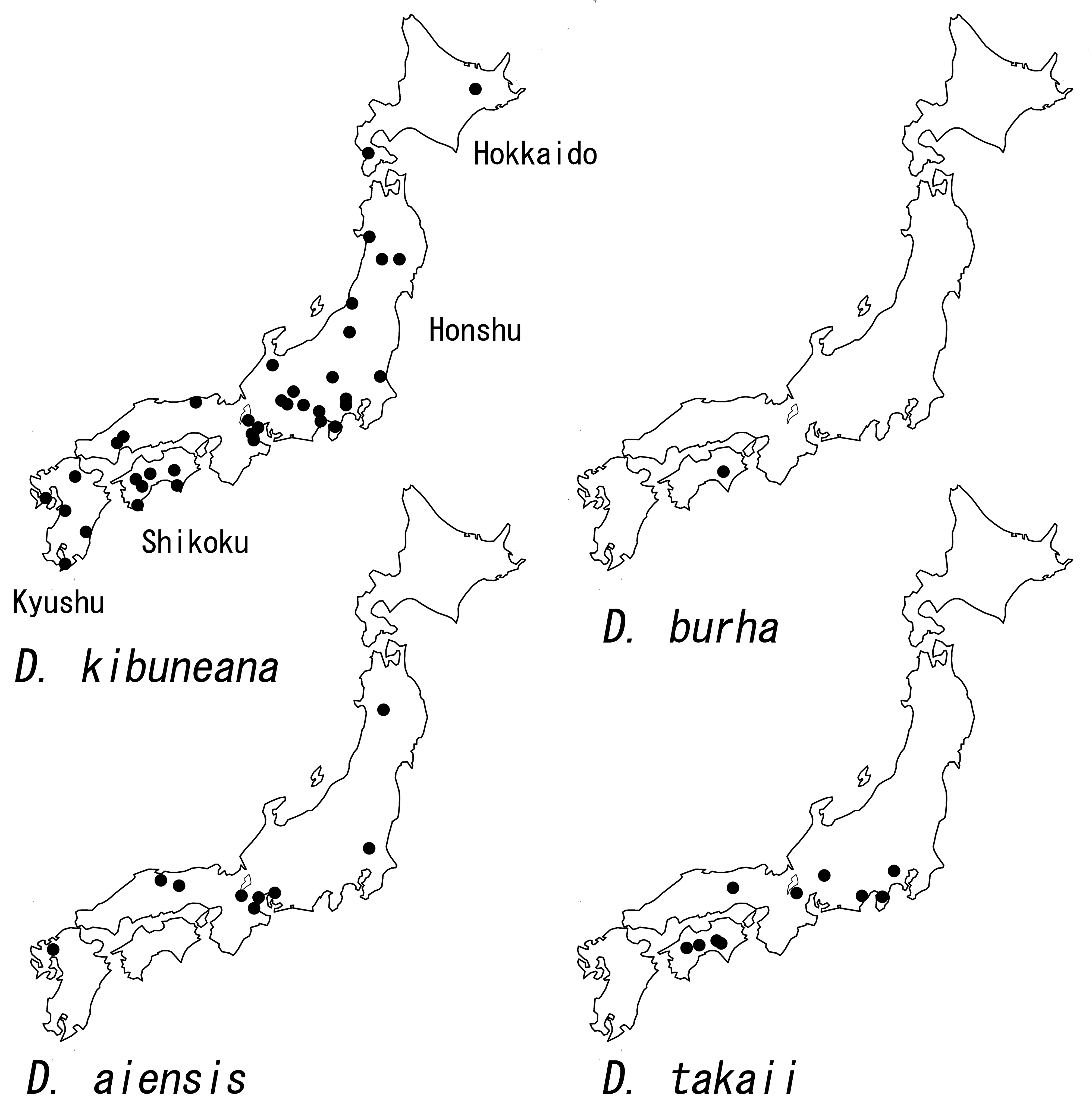
Distribution. Japan (Shikoku, Ryukyu islands), Pakistan, India, Nepal, Thailand, Vietnam, Taiwan.
Japanese name. Buruha-miyama-shima-tobikera.
Remarks. The single male specimen available in this study has many characteristics which agree with those of D. burha described or redescribed by Schmid (1961), Malicky (2002), and Ito & Nozaki (2018); for example, the length of the lateral filaments of abdominal segment V, the size of the internal glands of segment V, lack of internal glands of segment VIII, and the shape of the genitalia. The shape of the mesocaudal lobes and lateral lobes of the segment X are different from those of males collected from Ryukyu islands, southwestern Japan ( Ito & Nozaki 2018), but similar to those illustrated in the original description ( Schmid 1961, pl. 17, figs 7, 8). Schmid (1961) and Malicky (2002) reported variations of the male genitalia in this species.
Ito, T. & Nozaki, T. (2018) The family Hydropsychidae Curtis (Trichoptera) in the Ryukyu Archipelago, southwestern Japan. Zootaxa, 4504 (4), 545 - 565. https: // doi. org / 10.11646 / zootaxa. 4504.4.6
Malicky, H. (2002) Ein Beitrag zur Kenntnis asiatischer Arten der Gattung Diplectrona Westwood 1840 (Trichoptera, Hydropsy chidae) (gleichzeitig Arbeit Nr. 34 uber thailandische Kocherfliegen). Linzer Biologische Beitrage, 34 (2), 1201 - 1236.
Schmid, F. (1961) Trichopteres du Pakistan. 4 me partie (fin). Tijdschrift voor Entomologie, 104, 187 - 230, pls. 13 - 25.
Sun, C-h. (2017) Eight new species of Diplectrona (Trichoptera: Hydropsychidae) from China. Journal of the Kansas Entomological Society, 90 (2), 146 - 161. https: // doi. org / 10.2317 / 0022 - 8567 - 90.2.146
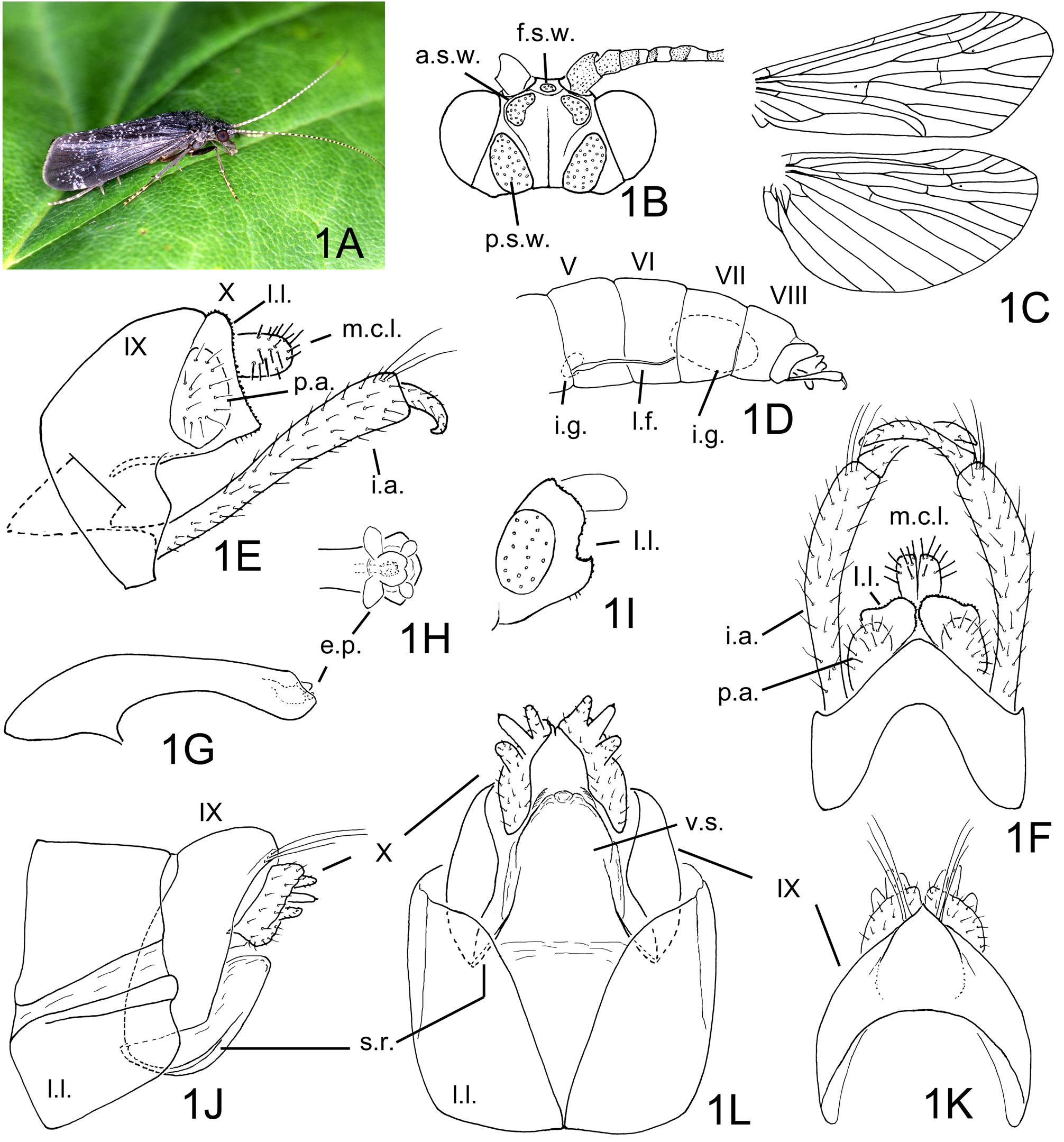
FIGURE 1. Diplectrona kibuneana Tsuda 1940. 1A–1I, male: 1A, habitus (Kochi, photographed by M. Takai), right lateral; 1B, head, dorsal; 1C, right wings, dorsal; 1D, abdominal segments V–X, left lateral; 1E, genitalia, left lateral; 1F, same, dorsal; 1G, phallic apparatus, left lateral; 1H, same, apical part, dorsal; 1I, segment X, left lateral, type series male of Hydropsyche difficultata Kobayashi. 1J–1L, female genitalia: 1J, left lateral; 1K, dorsal; 1L, ventral. Abbreviations: a.s.w. = anterior setal wart (paired), e.p. = endothecal process (paired), f.s.w. = frontal setal wart, i.a. = inferior appendage (paired), i.g. = internal gland (paired) of segment V or VIII, m.c.l. = mesocaudal lobe of segment X (paired), l.f. = lateral filament (paired), l.l. = lateral lobe of segment VIII in female or X in male (paired), p.a. = preanal appendage of segment X (paired), p.s.w. = posterior setal wart (paired), s.r. = sclerotized rib (paired), V–X = abdominal segments V–X, v.s. = vulval scale.
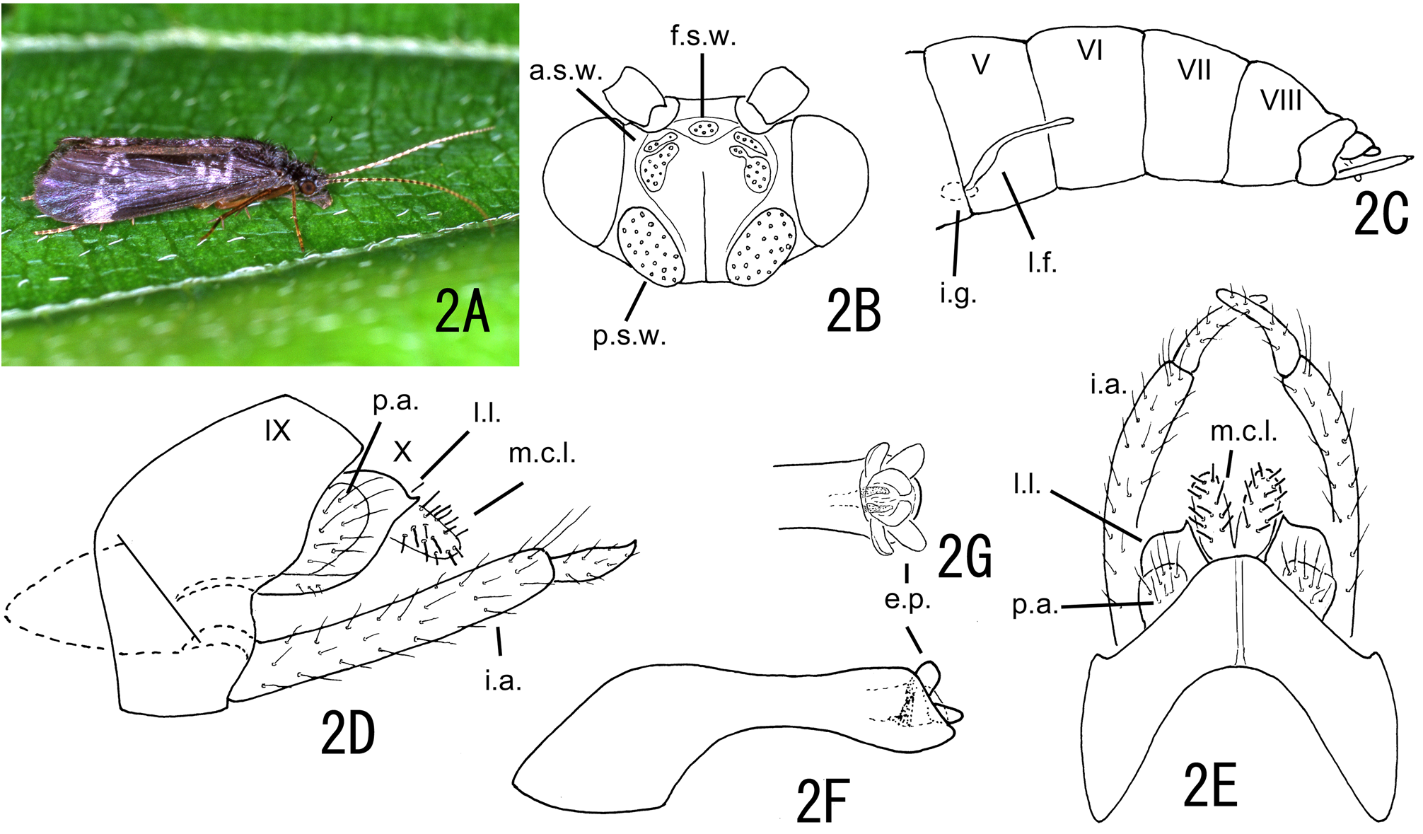
FIGURE 2. Diplectrona burha Schmid 1961. 2A–2G, male: 2A, habitus (Kochi, photographed by M. Takai), right lateral; 2B, head dorsal; 2C, abdominal segments V–X, left lateral; 2D, genitalia, left lateral; 2E, same, dorsal; 2F, phallic apparatus, left lateral; 2G, same, apical part, dorsal. Abbreviations: a.s.w. = anterior setal wart (paired), e.p. = endothecal process (paired), f.s.w. = frontal setal wart, i.a. = inferior appendage (paired), i.g. = internal gland (paired) of segment V, m.c.l. = mesocaudal lobe of segment X (paired), l.f. = lateral filament (paired), l.l. = lateral lobe of segment X (paired), p.a. = preanal appendage of segment X (paired), p.s.w. = posterior setal wart (paired), V–X = abdominal segments V–X.
No known copyright restrictions apply. See Agosti, D., Egloff, W., 2009. Taxonomic information exchange and copyright: the Plazi approach. BMC Research Notes 2009, 2:53 for further explanation.
|
Kingdom |
|
|
Phylum |
|
|
Class |
|
|
Order |
|
|
Family |
|
|
Genus |
Diplectrona burha Schmid 1961
| Nozaki, Takao 2021 |
Diplectrona burha
| Schmid 1961 |