
Micropora amapaensis Ramalho & Moraes, 2021
|
publication ID |
https://doi.org/10.11646/zootaxa.4950.1.1 |
|
publication LSID |
lsid:zoobank.org:pub:B9578A01-9B27-40B9-BEF9-C6DEB714C652 |
|
DOI |
https://doi.org/10.5281/zenodo.4643265 |
|
persistent identifier |
https://treatment.plazi.org/id/817C8781-FFEC-FFA8-A2E2-FA2AFE8AFD18 |
|
treatment provided by |
Plazi |
|
scientific name |
Micropora amapaensis Ramalho & Moraes |
| status |
sp. nov. |
Micropora amapaensis Ramalho & Moraes n. sp.
( Fig. 4A–C View FIGURE 4 )
Material examined. Holotype: MNRJBRY-1408 ; Paratype: MNRJBRY-1429: both from Brazil, Amapá state ( Sta #3, 03°35.4267’N – 049°07.6028’W), 90 m, on rhodoliths, 26 September 2014, collected by Fernando Moraes & Rodrigo Moura (NHo Cruzeiro do Sul ). GoogleMaps
Etymology. Referring to the type locality, Amapá state, Northern Brazil.
Diagnosis. Autozooids with paired, smooth tubercles lateral to orifice; oval to slit-like, asymmetrical opesiules, aligned with tubercles; proximal border of the opesia with a distinct straight and incomplete bar; avicularia absent; smooth, rounded triangular ovicell, closed by zooidal operculum.
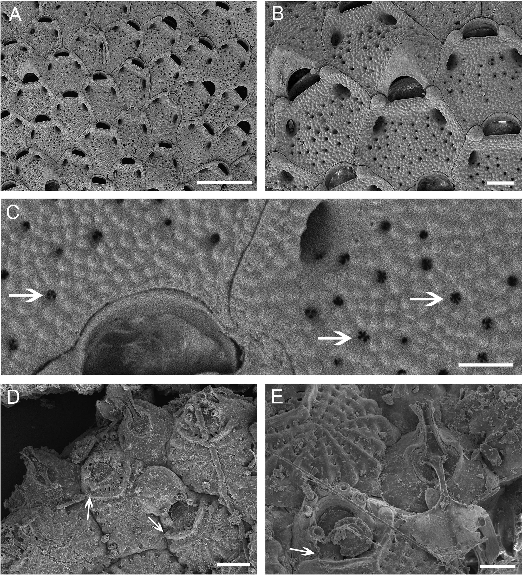
Description. Colony encrusting, multiserial, unilaminar. Autozooids disposed regularly in quincunx, separated by fine sutures, polygonal, slightly longer than wide [L 359–434–503 (SD 48, N 17); W 259–358– 421 µm (SD 38, N 20)], distally rounded ( Fig. 4A–B View FIGURE 4 ). Cryptocyst granular raised proximally to the opesia, perforated by small and scattered pseudopores ( Fig. 4B View FIGURE 4 ) with radial spines ( Fig. 4C View FIGURE 4 ). A pair of smooth tubercles located laterally to the orifice. Opesiules large, oval to slit-like [L 56–67– 85 µm (SD 7, N 20)], paired, asymmetrical in size and shape, aligned with the tubercles ( Fig. 4B View FIGURE 4 ). Opesia transversally D-shaped, wider than long [L 61–74–90 (SD 8, N 19); W 110–128– 143 µm (SD 8, N 19)], distal part smooth, proximal border with a distinct straight and incomplete bar not touching the proximal corners ( Fig. 4B View FIGURE 4 ). Ovicell wider than long [L 84–118– 137 µm (SD 24, N 4); Wmax (proximal width) 182–191– 201 µm (SD 8, N 4), Wmin (distal width) 60–85– 107 µm (SD 18, N 5)], smooth, rounded triangular, sometimes slightly raised distally, occupying part of the proximal region of the next distal zooid; aperture closed by zooidal operculum ( Fig. 4B View FIGURE 4 ). Avicularia absent.
Remarks. Three species of Micropora are known from Brazil: M. acuminata Winston, 2005 and M. angustiscapulis Winston et al., 2014 from Bahia state, and M. nodimagna Ramalho & Caliari, 2015 from Rio Grande do Sul state. The ovicell surface of M. acuminata is beaded with a sharp ridge; M. nodimagna has a very large orificial tubercle, smooth ovicell with a triangular, medially displaced suture, and several transverse lines on the proximal region; both M. angustiscapulis and M. amapaensis Ramalho & Moraes n. sp. lack avicularia, but the former species has smaller zooids (L 342–396– 450 µm, W 234–291– 360 µm), smaller orifice (L 45–51– 54 µm, W 90–101– 108 µm), raised, beaded cryptocyst edges, tubercles just below the proximal rim on either sides of the orifice, and opesiules narrower and shorter ( 5 µm). Winston et al. (2014) did not observe any complete ovicells in M. angustiscapulis . There are two other species of Micropora lacking interzooidal avicularia: M. coriacea inarmata Soule, 1959 , recorded from the Pacific Ocean, which has minute, knob-like tubercles, two small distal opesiules and ovicells lacking the triangular smooth calcification observed in M. amapaensis Ramalho & Moraes n. sp.; and M. mawatarii Arakawa, 2016 , described from Japan and with larger zooids (L 300–860 µm, W 230–720 µm), smooth gymnocyst, small tubercles and elliptical or circular small opesiules, contrasting with the large, oval to slit-like opesiules of the new species. Pseudopores with inner radiating denticulation seems to be a common character in Micropora , being mentioned in several species from different regions and geological ages (e.g., the Recent M. angustiscapulis from Brazil, M. mawatari and M. okadai Silén, 1942 both from Japan, and the Pliocene M. stellata Di Martino et al., 2019 from Florida).
No known copyright restrictions apply. See Agosti, D., Egloff, W., 2009. Taxonomic information exchange and copyright: the Plazi approach. BMC Research Notes 2009, 2:53 for further explanation.
|
Kingdom |
|
|
Phylum |
|
|
Class |
|
|
Order |
|
|
SubOrder |
Flustrina |
|
Family |
|
|
Genus |