
Homoneura (Homoneura) henanensis, Yang, Zhu & Hu, 1999
|
publication ID |
https://doi.org/10.11646/zootaxa.4365.3.5 |
|
publication LSID |
lsid:zoobank.org:pub:EA6D1CEF-0A4B-407C-9914-9F50A214696A |
|
persistent identifier |
https://treatment.plazi.org/id/2B0787BF-FF92-2925-FF08-FF46FE40F8FC |
|
treatment provided by |
Plazi (2019-05-20 15:39:58, last updated 2019-07-16 14:10:54) |
|
scientific name |
Homoneura (Homoneura) henanensis |
| status |
|
Key to separate the species in the Homoneura (Homoneura) henanensis group
[Modified from Shi & Yang, 2014, inserting the four new species into the key]
1. Wing with brown spot at tip of Sc and R 1 slightly elongating to costal margin (see Shatalkin, 2000: fig. 28); surstylus claviform with 3 long setulae, postgonite long coniform with 5 short setulae (see Sasakawa, 1982: fig. 4).................................................................................................... H. (H.) hirayamae (Matsumura)
- Wing without brown spot at tip of Sc and R 1 ................................................................ 2
2. Basal edge of brown apical spot on R 2+3 at same vertical level of crossvein dm-cu ( Figs 1 View FIGURES 1–4 , 34 View FIGURES 29–34 )......................... 3
- Basal edge of brown apical spot on R 2+3 behind vertical level of crossvein dm-cu ( Figs 13, 14 View FIGURES 10–14 , 20 View FIGURES 20–23 )...................... 6
3. Palpus entirely yellow ( Fig. 29 View FIGURES 29–34 ).......................................................................... 4
- Palpus yellow except for black at tip........................................... H. (H.) dadongshanica Shi & Yang
4. Subcostal cell hyaline; surstylus with a small acute teeth-like process in lateral view........... H. (H.) brevis Gao & Yang
- Subcostal cell brown apically ( Figs 1 View FIGURES 1–4 , 34 View FIGURES 29–34 ); surstylus without teeth-like process in lateral view......................... 5
5. Surstylus short and broad, with a row of long apical setulae ( Fig. 5 View FIGURES 5–9 ); pregonite blunt round with a medial setulae and postgonite subuliform apically ( Fig. 9 View FIGURES 5–9 )........................................................ H. (H.) jiangxiensis sp. nov.
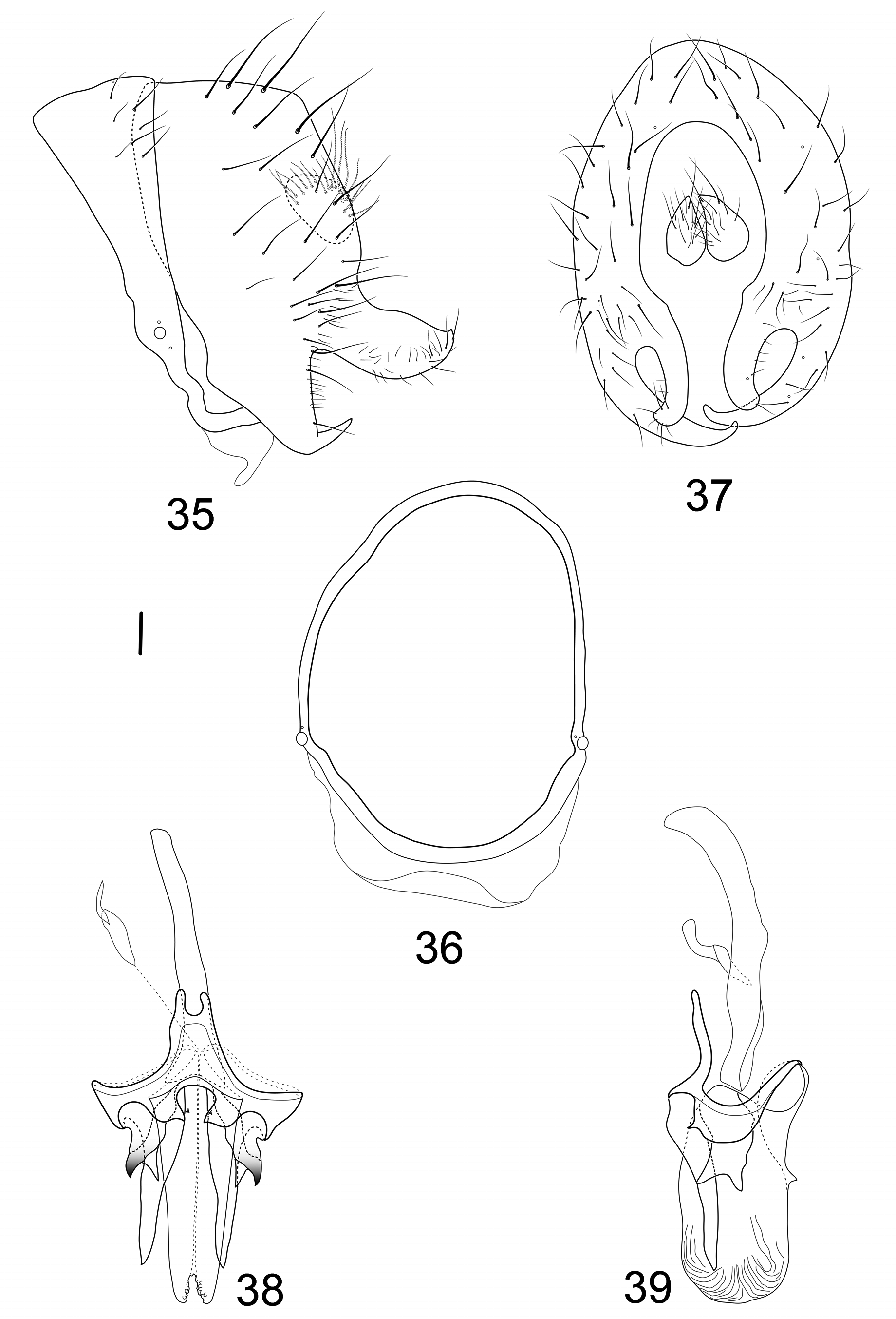
- Surstylus consisting of a wide knife-like process with many short ventral hairs and a triangular process, incurved apically ( Fig. 35 View FIGURES 35–39 ); pregonite subuliform and postgonite furcated apically ( Fig. 39 View FIGURES 35–39 )........................... H. (H.) stepheni sp. nov.
6. Basal edge of brown apical spot on R 2+3 at same vertical level of apical spot on R 4+5; apical spot on R 4+5 close to brown spot on crossvein dm-cu or at least 2/3 length of ultimate section of M 1 ................................................. 7
- Basal edge of brown apical spot on R 4+5 behind vertical level of apical spot on R 2+3; apical spot on R 4+5 far from brown spot on crossvein dm-cu and shorter than 2/3 length of ultimate section of M 1 ........................................... 10
7. Apical spot on R 4+5 close to brown spot on crossvein dm-cu; ctenidium with 16 short setae on fore femur; surstylus acute apically in lateral view; pregonite absent; postgonite consisting of a bifurcated process and a subuliform process in ventral view (see Shi & Yang, 2014: figs 102, 105)............................................. H. (H.) denticulata Shi & Yang
- Apical spot on R 4+5 about 2/3 length of ultimate section of M 1, not close to brown spot on crossvein dm-cu; ctenidium with 12- 14 short setae on fore femur; surstylus blunt apically in lateral view, pregonite with a reverse U-shaped process and postgonite consisting a pair of subuliform processes in ventral view...................................................... 8
8. Hypandrium with a short ventral process; pregonite with a pair of reverse U-shaped process in ventral view; postgonite short subuliform, but pregonite longer than postgonite in ventral view................................................ 9
- Hypandrium with a long ventral process; shape of pregonite and postgonite as above, but pregonite shorter than postgonite in ventral view (see Shi & Yang, 2014: figs 198, 199)............................ H. (H.) pseudograndis Papp & Gaimari
9. Phallus with a pair of lateral teeth subapically in ventral view; two arms of reverse U-shaped pregonite asymmetrical distinctly (see Shi & Yang, 2014: figs 258, 259)............................................ H. (H.) simigrandis Shi & Yang
- Phallus without a pair of lateral teeth subapically in ventral view; two arms of reverse U-shaped pregonite almost symmetrical in length (see Papp & Gaimari, 2013: fig. 14)............................................ H. (H.) grandis (Kertész)
10. Wing with brown string-like spot on R 2+3 and apical spots on R 4+5 and M 1; epandrium slender and surstylus acute apically with a long seta in lateral view....................................................... H. (H.) curvispina Gao & Yang
- Wing with round, elliptical or quadrate spot on R 2+3, R 4+5 and M 1, not as above; epandrium and surstylus not as above...... 11
11. Wing with brown apical spots on R 2+3, R 4+5 and M 1 entirely confluent, or slightly confluent and forming pale brown connecting area between apical spots on R 2+3, R 4+5 and M 1 .............................................................. 12
- Wing with brown apical spots on R 4+5 and M 1 confluent, separated from apical spot on R 2+3, or apical spots on R 2+3, R 4+5 and M 1 entirely separated..................................................................................... 15
12. Brown medial spot on R 4+5 separated from brown cloud on crossvein dm-cu...................................... 13
- Brown medial spot on R 4+5 confluent with brown cloud on crossvein dm-cu....................................... 14
13. Body length 8.6 mm; basal edge of brown apical spot on R 2+3 at same vertical level of crossvein dm-cu; a large rectangular spot on R 4+5, at middle point of distance between crossveins r-m and dm-cu; only female known....................................................................................................... H. (H.) yaromi Yang, Hu & Zhu
- Body length 5.7–6.3 mm; basal edge of brown apical spot on R 2+3 behind vertical level of crossvein dm-cu; a small round or quadrate spot on R 4+5, slightly beyond middle point of distance between crossveins r-m and dm-cu ( Figs 13, 14 View FIGURES 10–14 ); both female and male known.................................................................... H. (H.) martini sp. nov.
14. Antennal 1 st flagellomere brown except for yellow base; fore femur with 4 posteroventral setae; hypandrium circular; surstylus wide claviform with two apical setulae in posterior view.......................... H. (H.) guizhouensis Gao & Yang
- Antennal 1 st flagellomere entirely yellow; fore femur with 6 posteroventral setae; hypandrium semicircular; surstylus consisting of a triangular outer process and a short claviform inner process in lateral view.............. H. (H.) yangi Gao & Yang
15. Wing with brown apical spot on R 4+5 and M 1 slightly confluent and forming pale brown connecting area between two apical spots; apical spot on R 2+3 distinctly separated from apical spot on R 4+5 ........................................... 16
- Wing with brown apical spots on R 2+3, R 4+5 and M 1 entirely separated............................................ 25
16. Mesonotum with acrostichal setulae in 10 rows............................................................. 17
- Mesonotum with acrostichal setulae in 6–8 rows............................................................ 19
17. Subcostal cell brown apically; abdominal tergites 2–5 without pale brown posterior margin; postgonite extending to apical tip of phallus in ventral view (see Gao & Yang, 2004: fig. 42); surstylus not as above................................. 18
- Subcostal cell hyaline; abdominal tergites 2–5 with pale brown posterior margin; postgonite not extending to apical tip of phallus in ventral view; surstylus bulged claviform, with long setulae in lateral view (see Yang et al, 2001: figs 7–9).................................................................................... H. (H.) bispinalis Yang, Hu & Zhu
18. Surstylus T-shaped and rounded apically in lateral view (see Yang et al, 2003: fig. 29-801B).................................................................................................. H. (H.) fujianensis Yang, Zhu & Hu
- Surstylus short claviform in lateral view and curved upward apically in posterior view (see Gao & Yang, 2004: figs 38, 40).............................................................................. H. (H.) tianeensis Gao & Yang
19. Abdomen pale brown, tergites 1–6 with black posterior margin; surstylus straight claviform in lateral view.............................................................................................. H. (H.) serrata Gao & Yang
- Abdomen yellow, tergites 1–6 without brown posterior margin; surstylus not as above.............................. 20
20. Mid femur with 5 a ................................................................................... 21
- Mid femur with 4 a; surstylus not as above................................................................ 22
21. Surstylus consisting of a small acute apical process, directed downward and a slender knife-like process with dense setulae on dorsal margin in lateral view.................................................. H. (H.) henanensis Yang, Zhu & Hu
- Surstylus long claviform in lateral view with1–2 long setulae and a few of short hairs............. H. (H.) pangae sp. nov.
22. Wing with a brown spot between r-m and apical spot on R 4+5 distinctly or slightly confluent with brown spot on crossvein dmcu; epandrium not projecting backward, surstylus claviform or digitiform........................................ 23
- Wing with a brown quadrate spot between r-m and apical spot on R 4+5 separated from brown spot on crossvein dm-cu; epandrium and surstylus not as above........................................................................ 24
23. Ctenidium with 15 short setae on ventral margin of fore femur; surstylus absent; pregonite short, broad and acute apically in ventral view; postgonite consisting of a furcated process and a slender subuliform process in ventral view....................................................................................... H. (H.) curvispinosa Yang, Hu & Zhu
- Ctenidium with 13 short setae on ventral margin of fore femur; surstylus digitiform with long setulae in lateral view; pregonite and postgonite furcated apically, pregonite shorter than postgonite in ventral view......... H. (H.) zonalis Yang, Zhu & Hu
24. Fore femur with 3 posteroventral setae; epandrium blunt triangular apically; surstylus separated from epandrium and originated from anterior ventral corner of epandrium, with dense tiny setulae on apical 2/3 (Fig. 300)................................................................................................ H. (H.) tianjingshanica Shi & Yang
- Fore femur with 4 posteroventral setae; epandrium and surstylus fused, blunt round apically................................................................................................. H. (H.) tianmushana Yang, Hu & Zhu
25. Ctenidium with 17–19 short setae on ventral margin of fore femur; epandrium with a small acute process on anterior ventral corner; surstylus consisting of a small triangular anterior ventral process and a broad apical process with a tiny triangular apical tip and long setulae in lateral view (see Shi & Yang, 2014: figs 327, 329)............ H. (H.) zhangjiajiensis Shi & Yang
- Ctenidium with 12–15 short setae on ventral margin of fore femur; epandrium and surstylus not as above............... 26
26. Ctenidium with 15 short setae on ventral margin of fore femur; pregonite and postgonite long subuliform in ventral view.. 27
- Ctenidium with 12–13 short setae on ventral margin of fore femur; pregonite and postgonite not as above............... 28
27. Surstylus very broad ball-like with a triangular process apically in laterl view; hypandrium H-shaped; phallus acute subapically in lateral view (see Wang et al, 2012: figs 30–33)......................... H. (H.) kuankuoshuiensis Wang & Yang
- Surstylus narrow, acute apically in lateral view; hypandrium Y-shaped; phallus blunt round subapically in lateral view (see Shi & Yang, 2014: figs 66, 70).......................................................... H. (H.) chinensis Malloch
28. Subcostal cell hyaline apically; surstylus consisting of a slender knife-shaped process and a furcated process with several setulae on subapical and apical margin and a small tooth on lateral margin in lateral view (see Wang et al, 2012: fig. 18)........................................................................................ H. (H.) caoi Wang & Yang
- Subcostal cell dark apically; surstylus not as above.......................................................... 29
29. Mid femur with 5 a; mesonotum with acrostichal setulae in 8 rows; surstylus wide claviform, truncate apically, without acute apical process in lateral view; pregonite absent; postgonite wide triangular, constricted apically and curved upward in ventral view....................................................................... H. (H.) curvata Yang, Zhu & Hu
- Mid femur with 4 a or 6–8 a; mesonotum with acrostichal setulae in 10 rows.......................................... 30
30. A brown elliptical spot present between r-m and apical spot on R 4+5; mid femur with 6–8 a; surstylus curved knife-like, acute apically in lateral view; pregonite absent and postgonite longer than phallus, long subuliform, curved forward apically in lateral view (see Gao & Yang, 2004: figs 31, 32, 36).................................... H. (H.) longispina Gao & Yang
- A brown square spot present between r-m and apical spot on R 4+5; mid femur with 4 a; surstylus short, triangular and acutate apically, with several long setae on dorsal margin and a row of short setulae on ventral margin in lateral view; both pregonite and postgonite short subuliform, about half length of phallus in ventral view (see Yang et al, 1999: figs 4–6).. H. (H.) acutata Yang, Zhu & Hu
Gao, C. X. & Yang, D. (2004) A review of the genus Homoneura from Guangxi, China (Diptera: Lauxaniidae). Raffles Bulletin of Zoology, 52 (2), 351 - 364.
Papp, L. & Gaimari, S. D. (2013) The holotype of Homoneura grandis (Kertesz, 1915) with description of a new species from Taiwan (Diptera: Lauxaniidae). Acta Zoologica Academiae Scientiarum Hungaricae, 59 (1), 31 - 39.
Sasakawa, M. & Ikeuchi, S. (1982) A revision of the Japanese species of Homoneura (Homoneura) (Diptera: Lauxaniidae). Part 1. Kontyu, 50 (3), 477 - 499.
Shi, L. & Yang, D. (2014) Supplements to species groups of the subgenus Homoneura in China (Diptera: Lauxaniidae: Homoneura), with descriptions of twenty new species. Zootaxa, 3890 (1), 1 - 117. https: // doi. org / 10.11646 / zootaxa. 3890.1.1
Wang, J. C., Gao, C. X. & Yang, D. (2012) Three new species of the Homoneura (Homoneura) henanensis species group (Diptera, Lauxaniidae), with a key to Chinese species. Zootaxa, 3262, 35 - 45.
Yang, D, Zhu, F. & Hu, X. Y. (1999) New species of Lauxaniidae from Henan (Diptera: Acalyptratae). In: Shen, X. C. & Shi, Z. Y. (Eds.), Fauna and Taxonomy of Insects in Henan, Vol. 4. China Agricultural Scientech Press, Beijing, pp. 211 - 217.
Yang, D., Hu, X. Y. & Zhu, F. (2001) Diptera: Lauxaniidae. In: Wu, H. & Pan, C. W. (Eds.), Insects of Tianmushan National Nature Reserve. Science Press, Beijing, pp. 446 - 453.
Yang, D., Zhu, F. & Hu, X. Y. (2003) Diptera: Lauxaniidae. In: Huang, B. K. (Ed.), Fauna of Insects in Fujian Province of China, Vol. 8. Fujian Science and Technology Publishing House, Fuzhou, pp. 555 - 558.
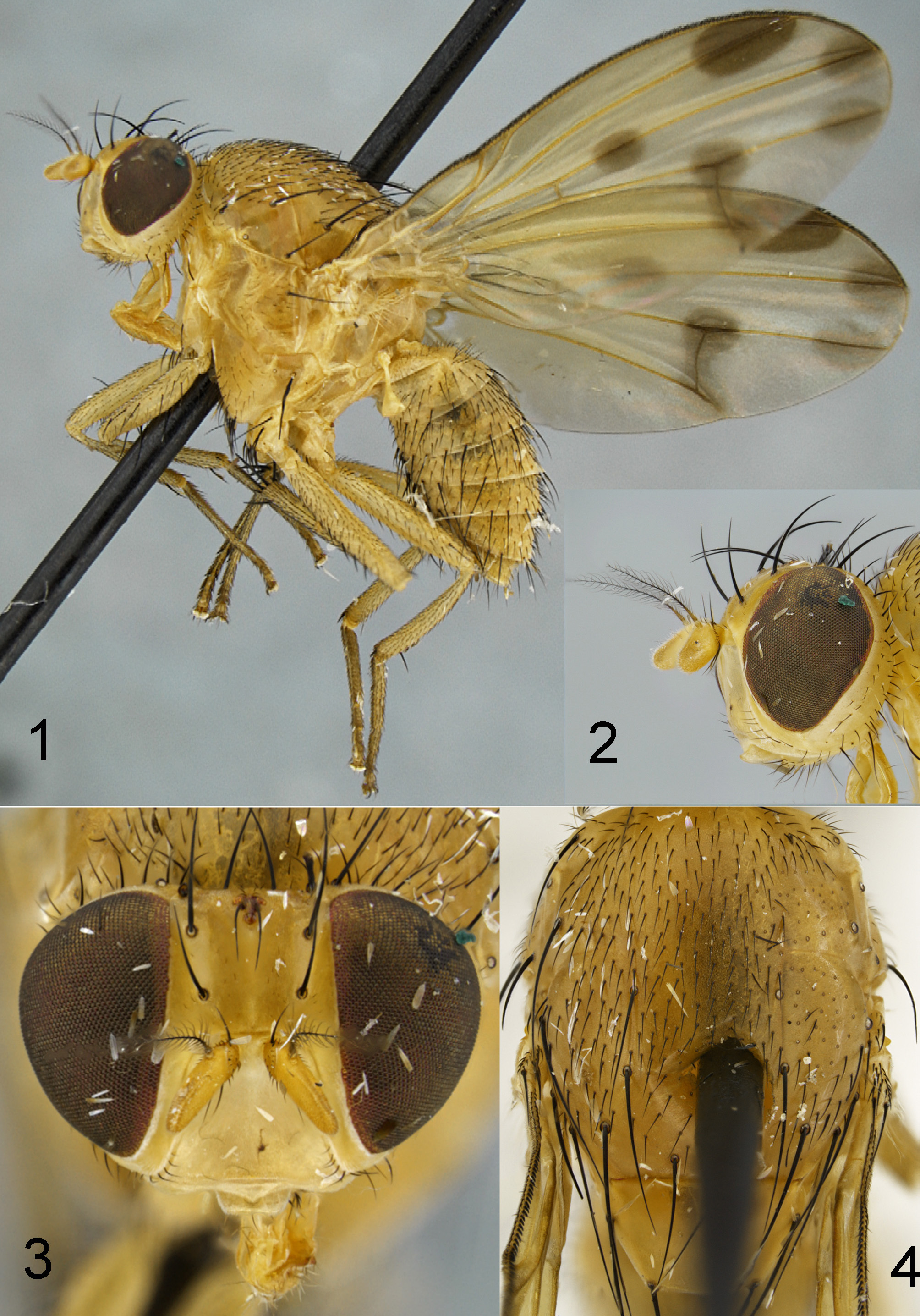
FIGURES 1–4. Homoneura (Homoneura) jiangxiensis sp. nov. Female. 1. body, lateral view; 2, 3. head, anterior and lateral view; 4. thorax, dorsal view.
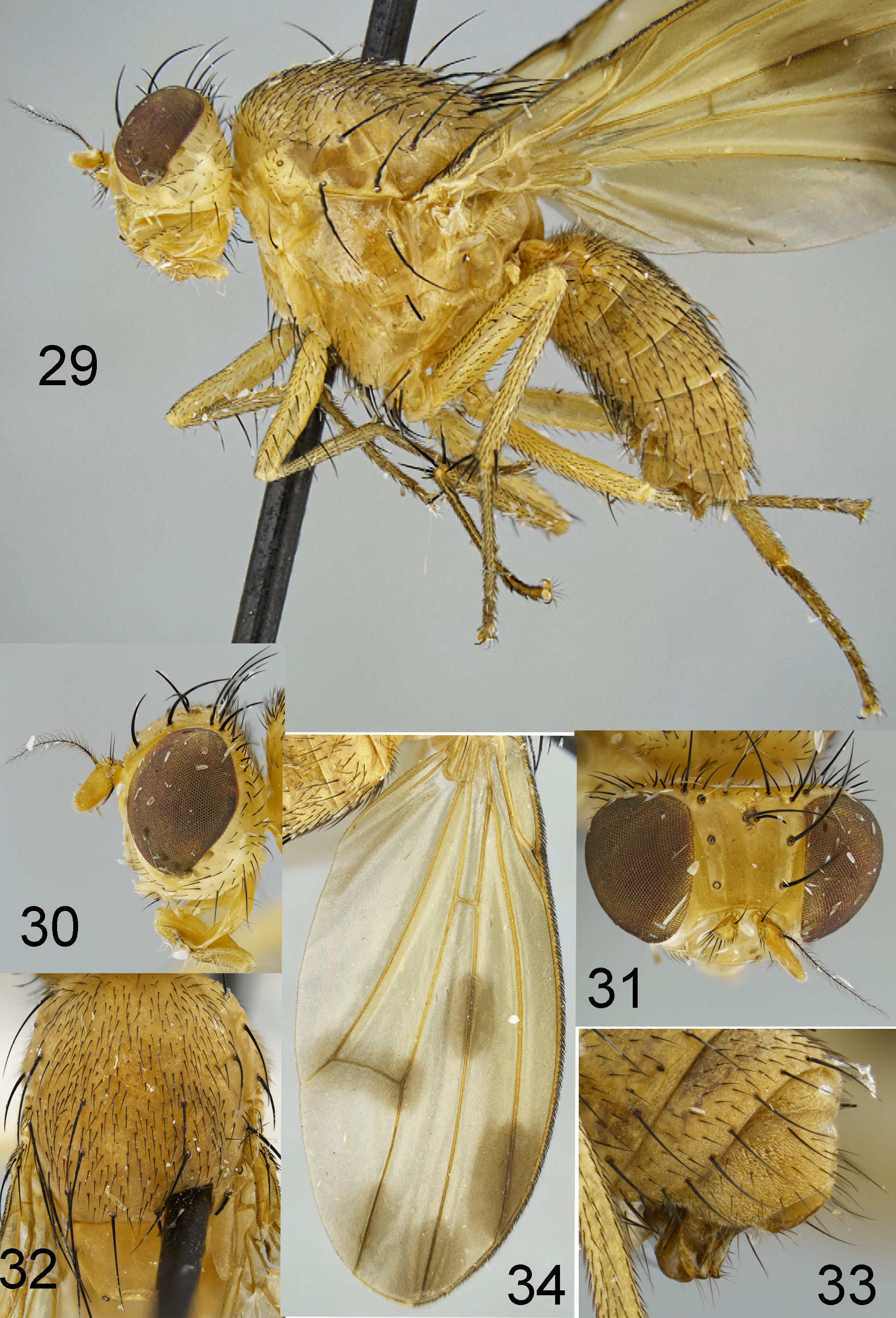
FIGURES 29–34. Homoneura (Homoneura) stepheni sp. nov. Female. 29. body, lateral view; 30, 31. head, lateral and dorsal view; 32. thorax, dorsal view; 33. apex of male abdomen, lateral view; 34. wing.
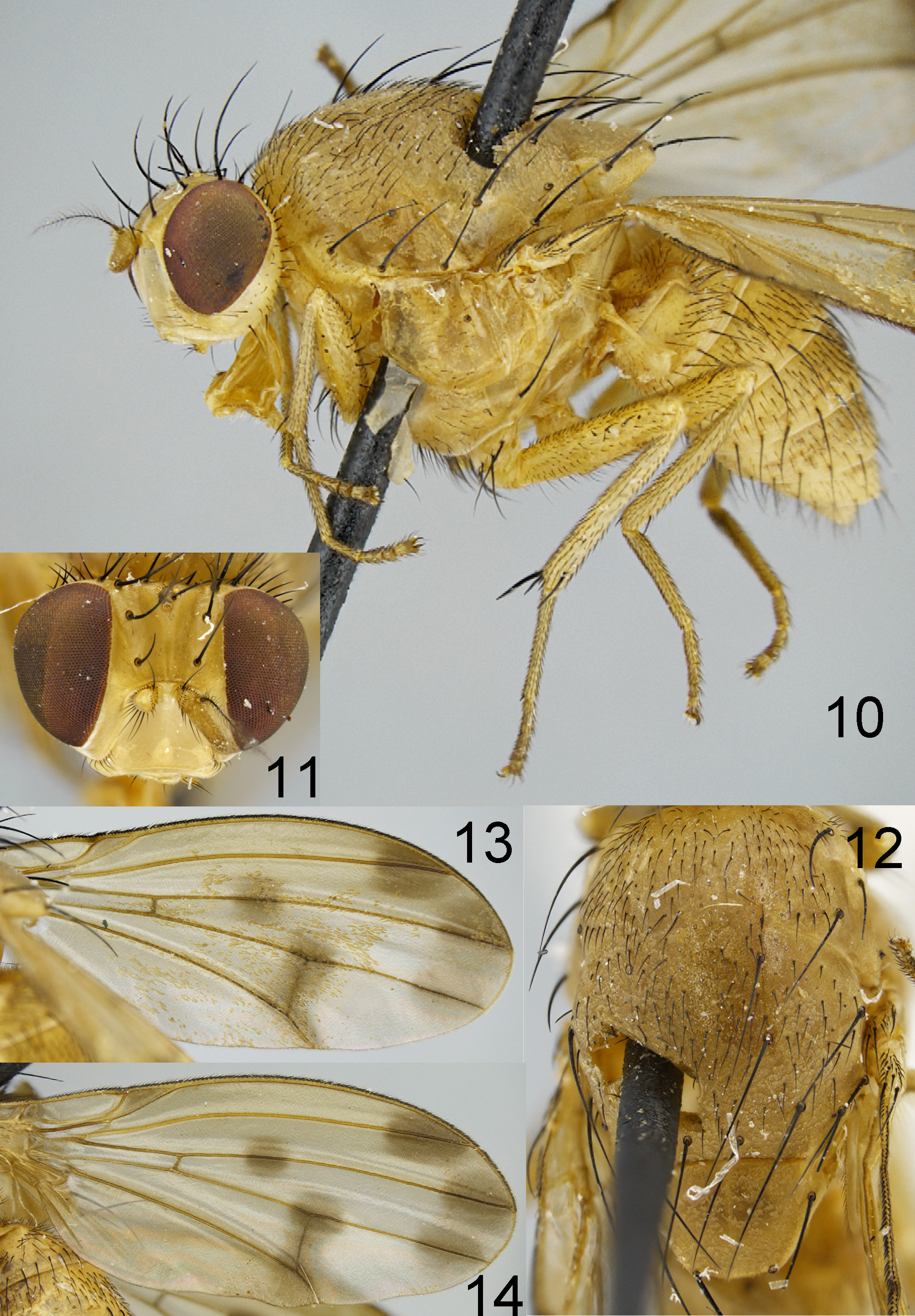
FIGURES 10–14. Homoneura (Homoneura) martini sp. nov. Female. 10. body, lateral view; 11. head, anterior view; 12. thorax, dorsal view; 13,14. wing.

FIGURES 20–23. Homoneura (Homoneura) pangae sp. nov. Male. 20. body, lateral view; 21. head, lateral view; 22. thorax, dorsal view; 23. apex of abdomen, lateral view.
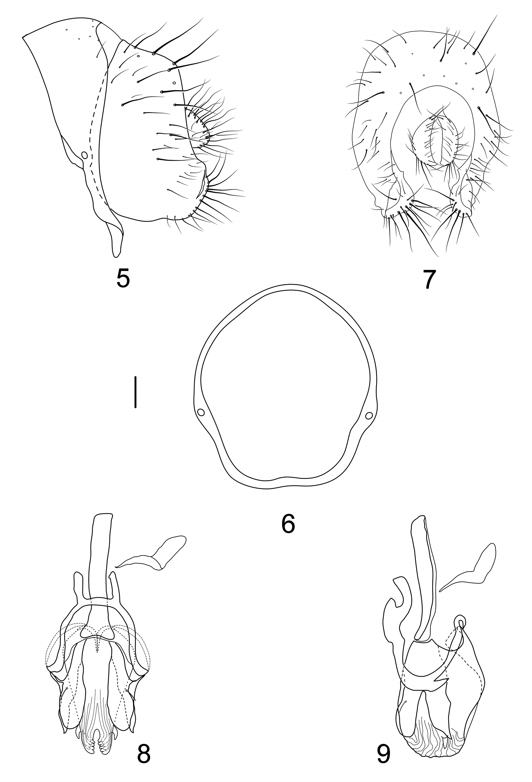
FIGURES 5–9. Homoneura (Homoneura) jiangxiensis sp. nov. Male. 5. syntergosternite and epandrium, lateral view; 6. syntergosternite, anterior view; 7. epandrial complex, posterior view; 8. phallus complex, ventral view; 9. phallus complex, lateral view. Scale bar =0.1mm.
No known copyright restrictions apply. See Agosti, D., Egloff, W., 2009. Taxonomic information exchange and copyright: the Plazi approach. BMC Research Notes 2009, 2:53 for further explanation.
|
Kingdom |
|
|
Phylum |
|
|
Class |
|
|
Order |
|
|
Family |
|
|
Genus |
1 (by plazi, 2019-05-20 15:39:58)
2 (by ImsDioSync, 2019-05-20 15:54:57)
3 (by ImsDioSync, 2019-05-20 15:55:51)
4 (by ImsDioSync, 2019-05-21 18:30:18)
5 (by ImsDioSync, 2019-07-16 14:10:54)
6 (by ImsDioSync, 2019-08-14 14:06:56)
7 (by ExternalLinkService, 2019-09-25 22:34:54)
8 (by ExternalLinkService, 2022-01-29 19:32:11)
9 (by ExternalLinkService, 2022-02-10 20:31:01)
10 (by plazi, 2023-10-30 14:41:12)