
Doriprismatica balut Matsuda and Gosliner, 2018
|
publication ID |
https://doi.org/ 10.11646/zootaxa.4444.5.1 |
|
publication LSID |
lsid:zoobank.org:pub:6A536780-96AE-42B0-913E-C05767BC63EC |
|
DOI |
https://doi.org/10.5281/zenodo.5981640 |
|
persistent identifier |
https://treatment.plazi.org/id/140387BF-486A-F00B-0D88-FA48FC42F841 |
|
treatment provided by |
Plazi |
|
scientific name |
Doriprismatica balut Matsuda and Gosliner |
| status |
sp. nov. |
Doriprismatica balut Matsuda and Gosliner View in CoL , sp. nov.
Figures (2H, 11A, B, 12, 13A–C)
Glossodoris sp. 1 Gosliner et al. 2008: p. 234, upper middle photo.
Doriprismatica sp. 1 Gosliner et al. 2015: p. 239, left middle photo.
Doriprismatica sp. A Matsuda & Gosliner 2017.
Type Material. Holotype: NMP 0 41278 (ex CASIZ-186096), one specimen 12 mm preserved, Philippines, Luzon, Batangas Province, Mabini (Calumpan Peninsula), Maricaban Strait, Murals dive site, coll: T.M. Gosliner, 3 May 2011, Hearst Philippine Biodiversity Expedition 2011, orig. fixative 95% EtOH. This specimen was tissue sampled (foot) for DNA analysis in Matsuda & Gosliner (2017), GenBank: KT600688 View Materials ( COI).
Paratypes: CASIZ-186095, one specimen, dissected, 10 mm preserved, Philippines, Batangas Province, Maricaban Island, Sepok Point, coll: M. Burke, 25 May 2011, Hearst Philippine Biodiversity Expedition 2011, orig. fixative 95% EtOH. This specimen was tissue sampled (foot) for DNA analysis in Matsuda & Gosliner (2017). CASIZ-105745A and 105745B, two specimens, dissected, (A) 13 mm preserved (B) 10.5 mm preserved, Philippines, Mindoro Island, North side of the island, mouth of the northwest passage, coll: R. McPeak, 28 February 1995, orig. fixative Bouin’s solution. CASIZ-182787, one specimen 8 mm preserved, Philippines, Batangas Province, Maricaban Island, Caban Island, Layag Layag, coll: T.M. Gosliner, 18 May 2010, Philippines Expedition 2010, orig. fixative 95% EtOH. CASIZ-208500, two specimens, Philippines, Mindoro: Oriental Mindoro Province, Puerto Galera, Palangan Point, coll: T.M. Gosliner, 0 7 April 2015, 2015 Verde Island Passage Expedition, orig. fixative 95% EtOH.
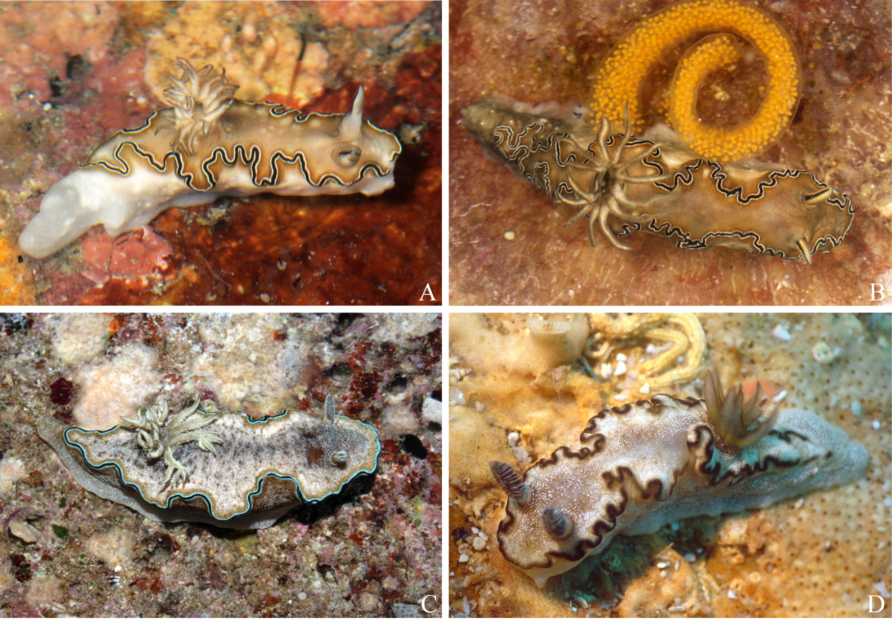
Etymology. Doriprismatica balut is named after the Filipino food, “ balut ”, which is a boiled duck egg that contains a developed embryo, to represent the presumed direct development of D. balut , based on its large egg size ( Fig. 11B View FIGURE 11 ).
Distribution. Specimens analyzed here have only been identified in the Philippines, although it has also been recorded from Indonesia ( Gosliner et al. 2008).
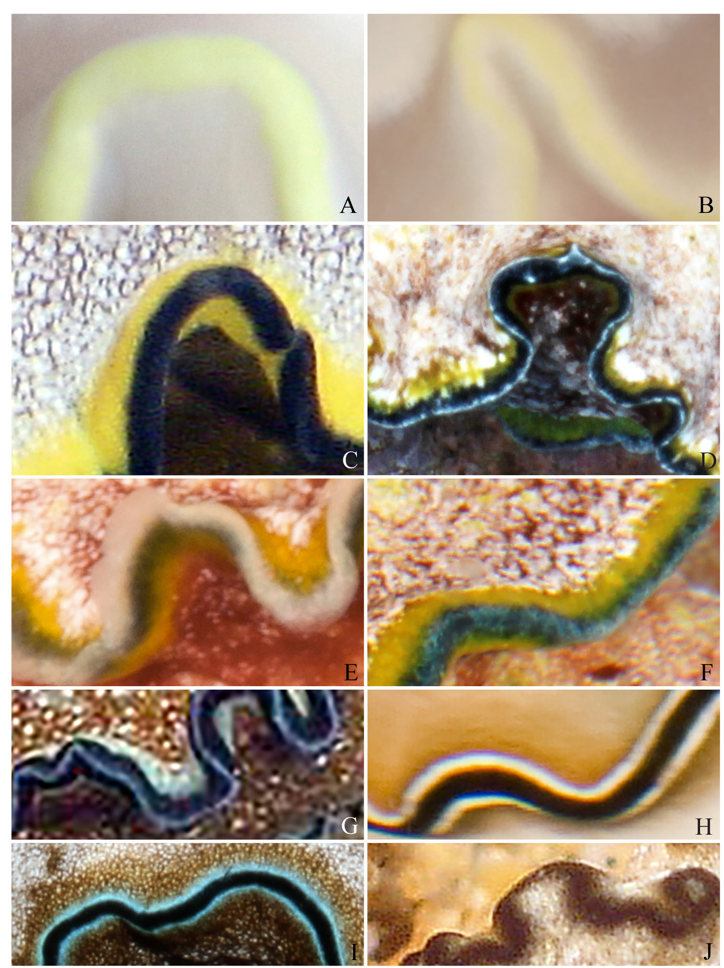
External morphology. Doriprismatica balut has an elongate body with a light caramel colored mantle that is slightly darker near the edge ( Figs. 11A, B View FIGURE 11 ). The mantle is surrounded by permanent and smaller semi-permanent undulations, and a series of marginal bands: a thin white inner band, followed by a thicker black one ( Fig. 2H View FIGURE 2 ). A second inner white band is found on the ventral surface of the mantle. The caramel color is present on the underside of the mantle, but the foot itself is semi-transparent white. The gill has 12–14 unipinnate lamellae arranged in a semicircle. A few gill branches are bifid near the apices. The gill sits a little more than half way back on the top of the mantle and surrounds the anus in an arc that opens posteriorly. The gill lamellae are the same color as the mantle on the underside and fades to a cream color facing inwards. The tips are outlined in black. The rhinophores are cream and caramel colors with a black vertical line running up the anterior and posterior sides. The rhinophores have 10–12 crowded lamellae. The genital opening is on the right side located slightly behind the rhinophores.
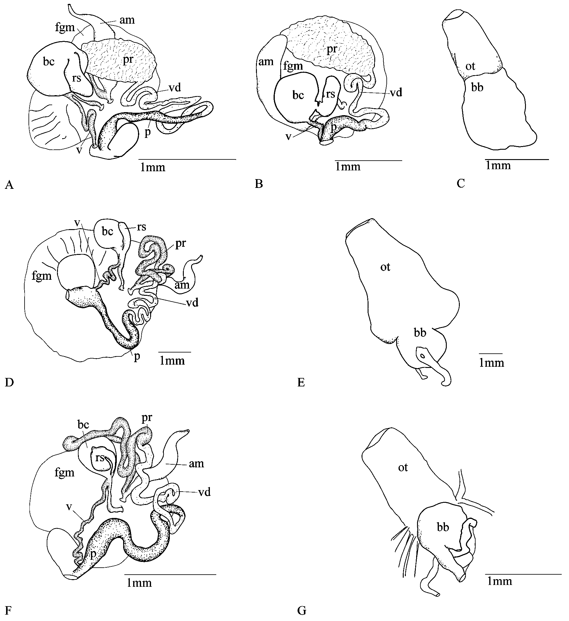
Internal anatomy. Radular structure ( Fig 12 View FIGURE 12 ). The muscular portion of the buccal mass is slightly longer than the oral tube ( Fig. 13C View FIGURE 13 ). The radula ribbon is long and wide ( Fig. 12D View FIGURE 12 ) (13 mm specimen 119 x 30.1. 30, 10 mm specimen 136 x 30.1.30). The rachidian tooth ( Fig. 12A View FIGURE 12 ) is reduced and is approximately a quarter of the length of the first lateral tooth. The first lateral tooth has 3–6 well defined large denticles flat against both sides of a longer central cusp. The remaining inner laterals are similar in appearance but do not have denticles on the inner side. The mid-laterals ( Fig. 12B View FIGURE 12 ) have a longer central cusp and the outer laterals are reduced and smooth ( Fig. 12C View FIGURE 12 ). The jaw rodlets are curved with a bifid tip ( Fig. 12E View FIGURE 12 ).
Reproductive system ( Figs. 13A, B View FIGURE 13 ). The vagina is short and connects to the receptaculum seminis duct just below the bursa copulatrix and receptaculum seminis sac, which is significantly smaller than the bursa copulatrix. The penial bulb is of medium length and connects to a short muscular vas deferens and long and folded prostate gland that is pressed to the female gland mass. The ampulla connects to the prostate gland before connecting to the female gland mass. The egg mass is translucent and laid in a spiral with bright yellow eggs, the size of which suggests this species may have direct development ( Fig. 11B View FIGURE 11 ).
Remarks. Doriprismatica balut is morphologically and molecularly distinct from the other Doriprismatica . Doriprismatica balut shares similarities with D. atromarginata ( Cuvier 1804) in its shape and body color, however D. balut is easily distinguished by the thick continuous black line around the mantle edge, where D. atromarginata’s black mantle band has multiple breaks. Doriprismatica balut also has an inner white line that is absent in D. atromarginata . The gill and rhinophores of D. atromarginata are black, whereas in D. balut they are the same color as the mantle and rhinophores and only have black bands running up the anterior and posterior edge. Doriprismatica balut also has a rachidian tooth, which is lacking in D. rossi sp. nov., D. atromarginata , Doriprismatica sibogae ( Bergh 1905) , and Doriprismatica paladentata ( Rudman 1986) . In D. paledentata , the inner lateral tooth is much broader whereas the remaining lateral teeth are narrow and elongate ( Rudman 1986: fig. 9). Doriprismatica stellata ( Rudman 1986) has a rachidian tooth, however the denticles are much larger and welldefined ( Rudman 1986: fig. 12) than in D. balut . Dorioprismatica marinae also has a rachidian row of teeth, but they are broad rather than triangular and have many denticles (Fig. 124).
There are also differences in internal anatomy. The vagina of D. balut is closer in length to Doriprismatica rossi and D. marinae , and shorter than D. atromarginata .
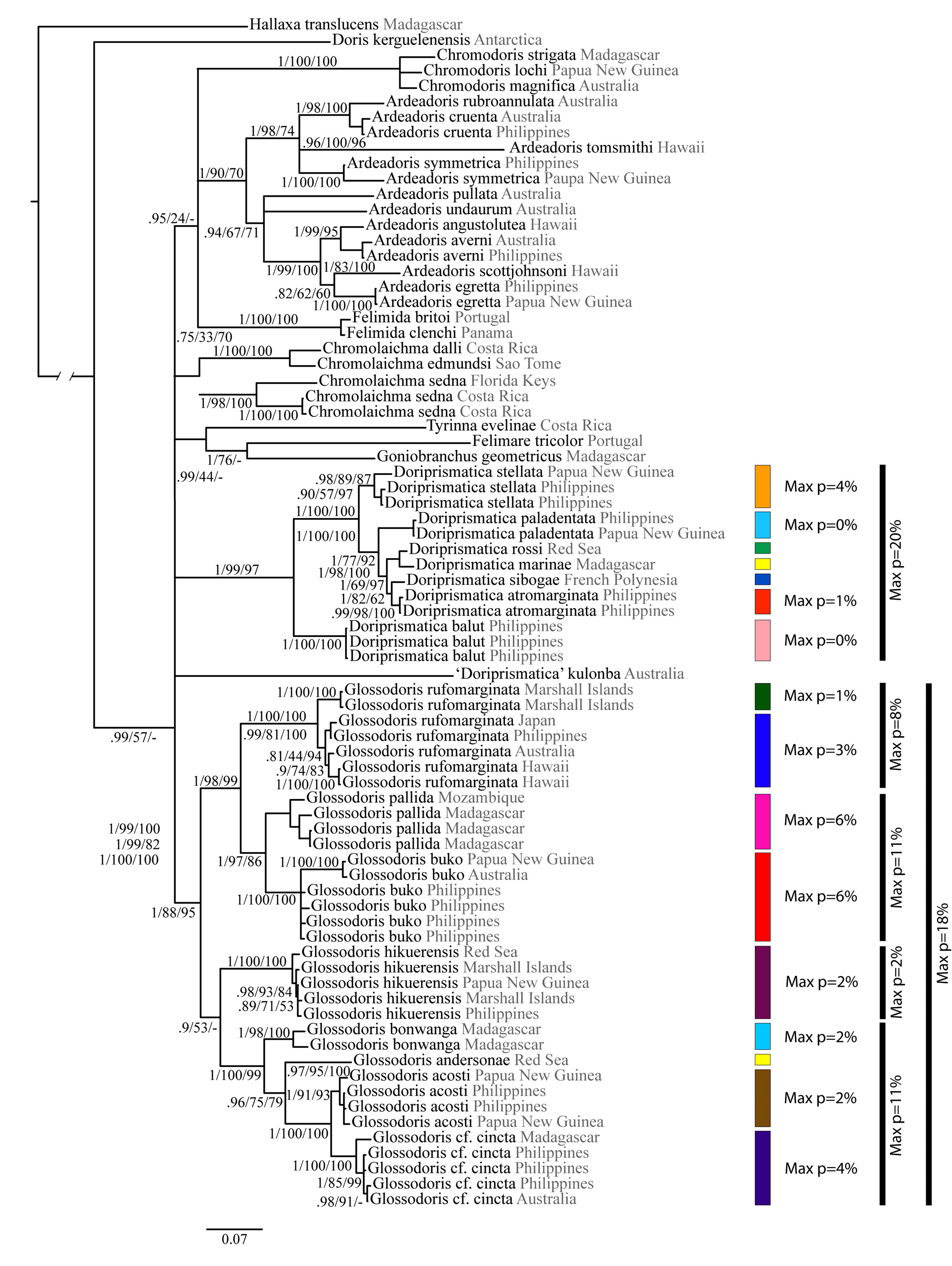
Doriprismatica balut is sister to the clade containing other Doriprismatica . Our ABGD analysis clearly shows the distinctness of this species, with no genetic variation found in the COI sequences in the three Philippine specimens examined by Matsuda & Gosliner (2017) ( Fig. 5 View FIGURE 5 ). The interspecific p-distances range from 11–16% between g D. balut and the other Doriprismatica that were studied.

No known copyright restrictions apply. See Agosti, D., Egloff, W., 2009. Taxonomic information exchange and copyright: the Plazi approach. BMC Research Notes 2009, 2:53 for further explanation.
|
Kingdom |
|
|
Phylum |
|
|
Class |
|
|
Order |
|
|
Family |
|
|
Genus |