
Psolidium spinuliferus (H. L. Clark)
|
publication ID |
https://doi.org/10.24199/j.mmv.2008.65.2 |
|
DOI |
https://doi.org/10.5281/zenodo.8070648 |
|
persistent identifier |
https://treatment.plazi.org/id/03FC87C9-5609-FFB3-A2AD-FADDFB06F0C9 |
|
treatment provided by |
Felipe (2023-06-21 13:59:15, last updated 2023-06-22 15:31:23) |
|
scientific name |
Psolidium spinuliferus (H. L. Clark) |
| status |
|
Psolidium spinuliferus (H. L. Clark) View in CoL
Table 1 View Table 1 , Figures 4a View Figure 4 , 8f View Figure 8 , 9a–d View Figure 9
Psolus spinuliferus H. L. Clark, 1938: 509–11 View in CoL , fig. 53.—H. L. Clark, 1946: 414.— Cannon and Silver, 1987: 29.—Rowe (in Rowe and Gates), 1995: 319.
Material examined. Northern Territory, Darwin Harbour, North Shell I, 12°29'48"S 130°53'12"E, coral rubble covered with sponges and some algae, 5 m, P.A.Hutchings, 16 Jul 1993, stn NT 346, AM J24096 (1) GoogleMaps . Western Australia, Perth, Cottesloe, Mudurup Rocks, c 70 m S of groyne, reef flat, Sargassum zone, on reef flat under thin veneer of sand , 31°59'51"S 115°45'01"E, 0–1 m, J. Keesing, 6 Feb 2007, WAM Z37479 About WAM (1); GoogleMaps Trigg I, c 100 m N of ‘island', inshore mixed algal zone mid-platform with thin veneer of sand overlaying reef , 31°52'29"S 115°45'04"E, 0–1 m, J. Keesing, 19 Feb 2007, Z37478 (1); GoogleMaps Waterman, Sargassum zone, mid-platform , 31°51'15"S 115°45'05"E, 0–1 m, J. Keesing, 14 Feb 2007, Z37468 (5); GoogleMaps from mixed localities, Cottesloe and Trigg I, inter-tidal platforms, on reef flat under thin veneer of sand , J. Keesing, Feb 2007, Z37469 (1) GoogleMaps .
Description. Psolidium species up to 20 mm long (preserved); dorsal and lateral body scales thin, single-layered, with spires, scales up to 1.5 mm wide; tube feet dorsally and laterally pass through scales, not conspicuous amongst spires.
Sole: peripheral band of tube feet, outer single series of smaller tube feet, inner single to zig-zag series; mid-ventral radial series irregular, double to zig-zag to scattered.
Dorsal and lateral ossicles: single-layered, thick, perforated plates (scales), irregularly oval, some with secondary thickening, most with vertical digitiform spire near margin; spires up to 400 μ m long, 120 μ m diameter, distally spinous.
Sole ossicles: knobbed plates, numerous, predominantly regular 4-holed thin plates, smooth to finely knobbed marginally, typically 80 μ m long; lacking cupped crosses, cups, rosettes.
Tentacle ossicles include: thick perforated plates, elongate, variable form, some with secondary layer development, up to 352 μ m long; numerous rosettes, large to small, frequently with 4 central perforations, densely branched, oval to elongate and distally rounded, up to 160 μ m long, intergrade with elongate plates.
Colour(live and preserved).White, dorsally and ventrally.
Distribution. Northern Territory (Darwin), to Western Australia (Perth); 0– 22 m.
Remarks. The dorsal and lateral tube feet are not conspicuous, and were not noticed by H. L. Clark (1938). Psolus spinuliferus H. L. Clark, 1938 is reassigned here to Psolidium Ludwig. The distinguishing characters of Psolidium spinuliferus (H. L. Clark) are the predominantly single-layered scales with vertical digitiform marginal spire. The type specimen ( MCZ no. 1669) was taken off the Eighty Mile Beach near Broome in northwestern Australia, at 18– 22 m.
Cannon, L. R. G., and Silver, H. 1987. Sea Cucumbers of Northern Australia. Queensland Museum: Brisbane, Australia. i - viii, 60 pp.
Clark, H. L. 1938. Echinoderms fromAustralia. An account of collections made in 1929 and 1932. Memoirs of the Museum of Comparative Zoology at Harvard College 55: 1 - 596, 28 pls, 63 figs.
Clark, H. L. 1946. The echinoderm fauna of Australia. Its composition and its origin. Carnegie Institution of Washington Publication 566: 1 - 567.
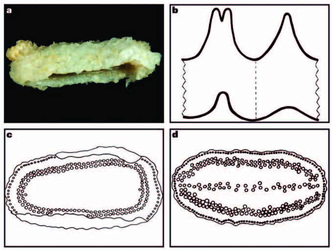
Figure 4. a, P. spinuliferus (H. L. Clark, 1938), Darwin (lateral view; 10 mm long; AM J24096; photo by L. Altoff); b, generalised form of radial (left) and interradial plates of the calcareous ring of Psolidium species (drawing by M. O’Loughlin); c–d, drawings of sole showing distribution of tube feet (by D. Maric); c, P. granuliferum H. L. Clark, 1938 (SAM K2176); d, P. karenae sp. nov. (SAM K2188).
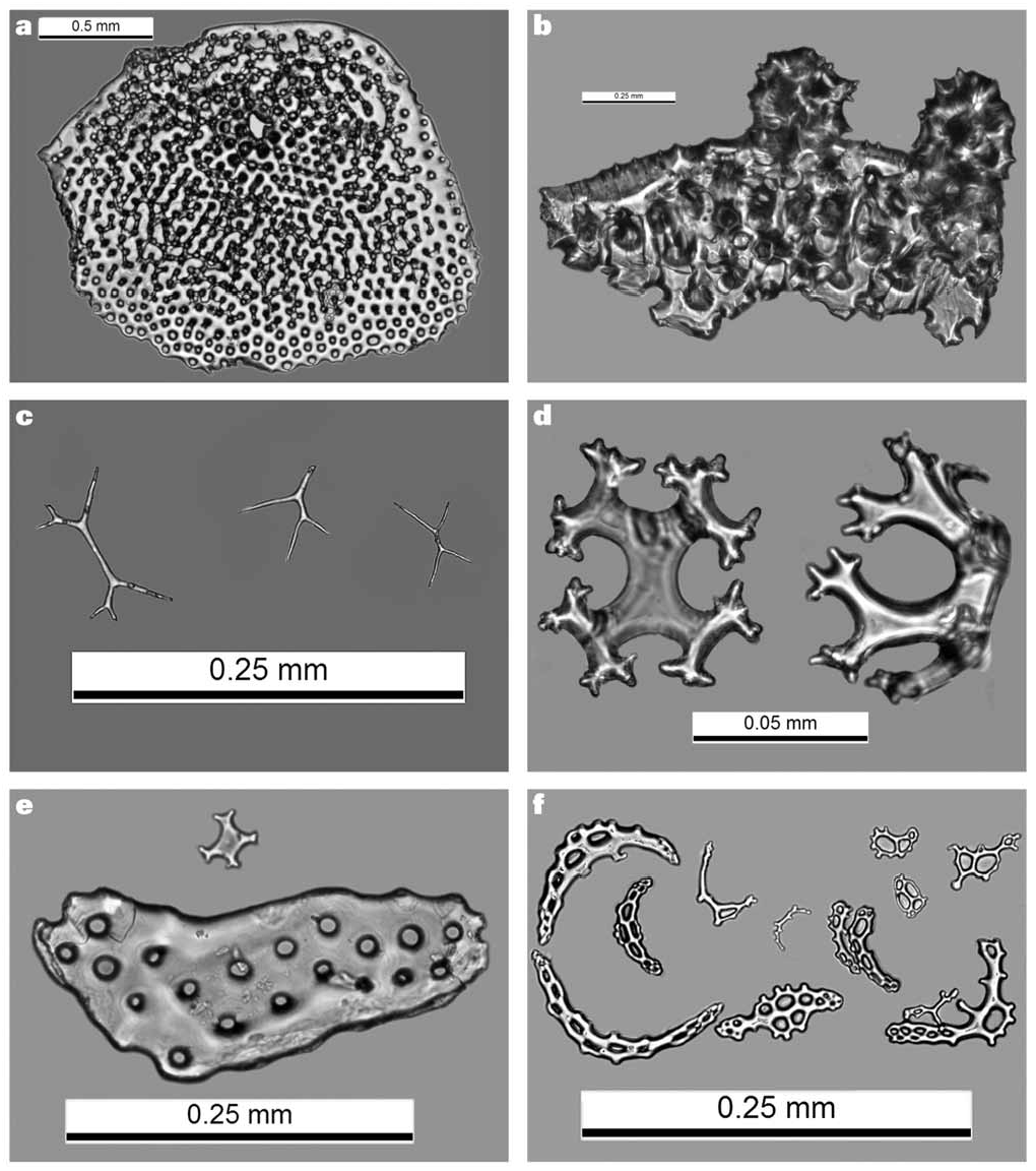
Figure 8. Photos of ossicles from Australian species of Psolidium Ludwig, 1886 (by Mark O’Loughlin and Chris Rowley): a–c, P. parmatus (Sluiter, 1901); a, dorsal scale with tube foot canal (holotype V.ECH.H1300); b, pillars on edge of part of scale (NMV F109378); c, dorsal “thorns” (holotype V.ECH.H1300); d–e, P. ravum Hickman, 1962 (SAM K2180); d, dorsal cupped crosses; e, cupped cross and plate from sole; f, P. spinuliferus (H. L. Clark, 1938), tentacle rods and dendritic branch endplates (top right) (AM J24096).
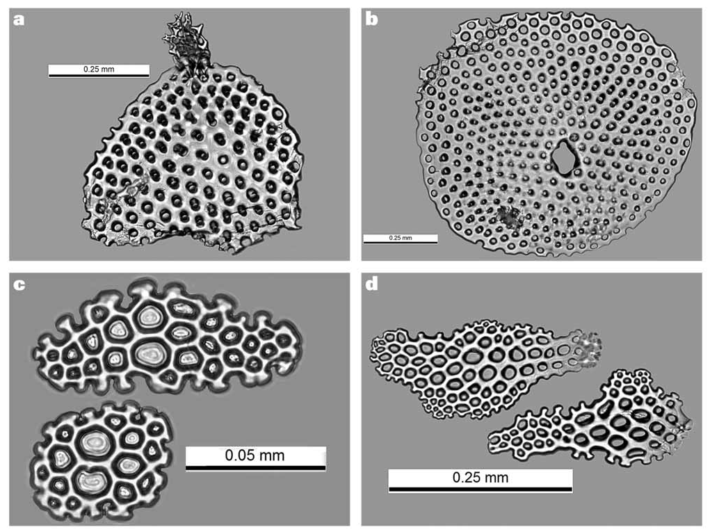
Figure 9. Photos of ossicles from Australian species of Psolidium Ludwig, 1886 (by Mark O’Loughlin and Chris Rowley): a–d, P. spinuliferus (H. L. Clark, 1938) (WAM Z37468); a, part of dorsal scale with marginal vertical spire; b, dorsal scale with tube foot canal and base of lost spire; c, tentacle rosettes; d, tentacle plates.
No known copyright restrictions apply. See Agosti, D., Egloff, W., 2009. Taxonomic information exchange and copyright: the Plazi approach. BMC Research Notes 2009, 2:53 for further explanation.
|
Kingdom |
|
|
Phylum |
|
|
Class |
|
|
Order |
|
|
Family |
|
|
Genus |
Psolidium spinuliferus (H. L. Clark)
| O’Loughlin, P. Mark & Ahearn, Cynthia 2008 |
Psolus spinuliferus H. L. Clark, 1938: 509–11
| Cannon, L. R. G. & Silver, H. 1987: 29 |
| Clark, H. L. 1946: 414 |
| Clark, H. L. 1938: 11 |