
Triconia onnuri, Cho & Böttger-Schnack & Kim & Lee, 2019
|
publication ID |
https://doi.org/10.5252/zoosystema2019v41a28 |
|
publication LSID |
urn:lsid:zoobank.org:pub:EB5117B3-49C3-4F42-B8A7-65C9F3841A5A |
|
DOI |
https://doi.org/10.5281/zenodo.4439443 |
|
persistent identifier |
https://treatment.plazi.org/id/515ECE8F-6AAE-47DD-AF7B-55D899441DC7 |
|
taxon LSID |
lsid:zoobank.org:act:515ECE8F-6AAE-47DD-AF7B-55D899441DC7 |
|
treatment provided by |
Felipe |
|
scientific name |
Triconia onnuri |
| status |
n. sp. |
Triconia onnuri n. sp.
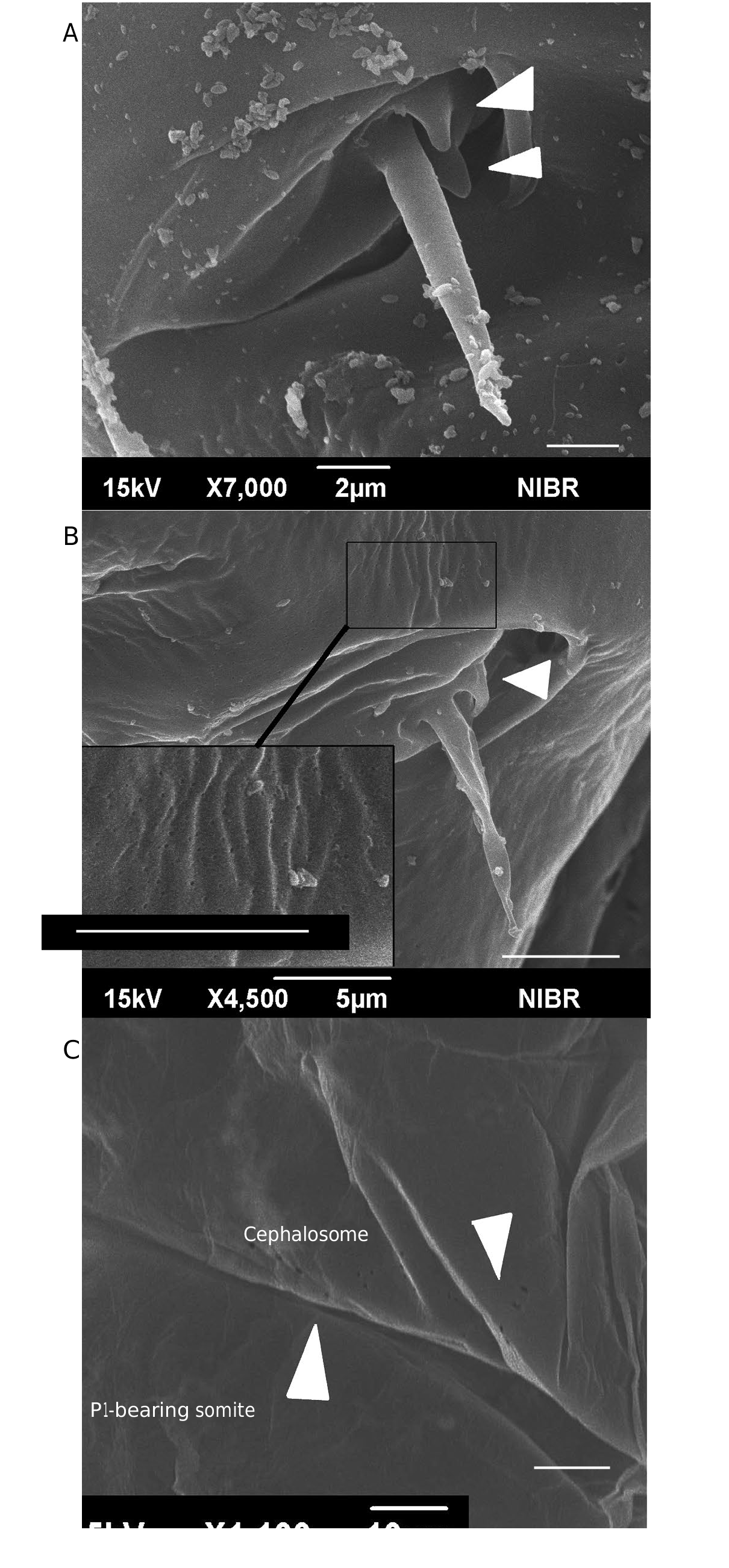
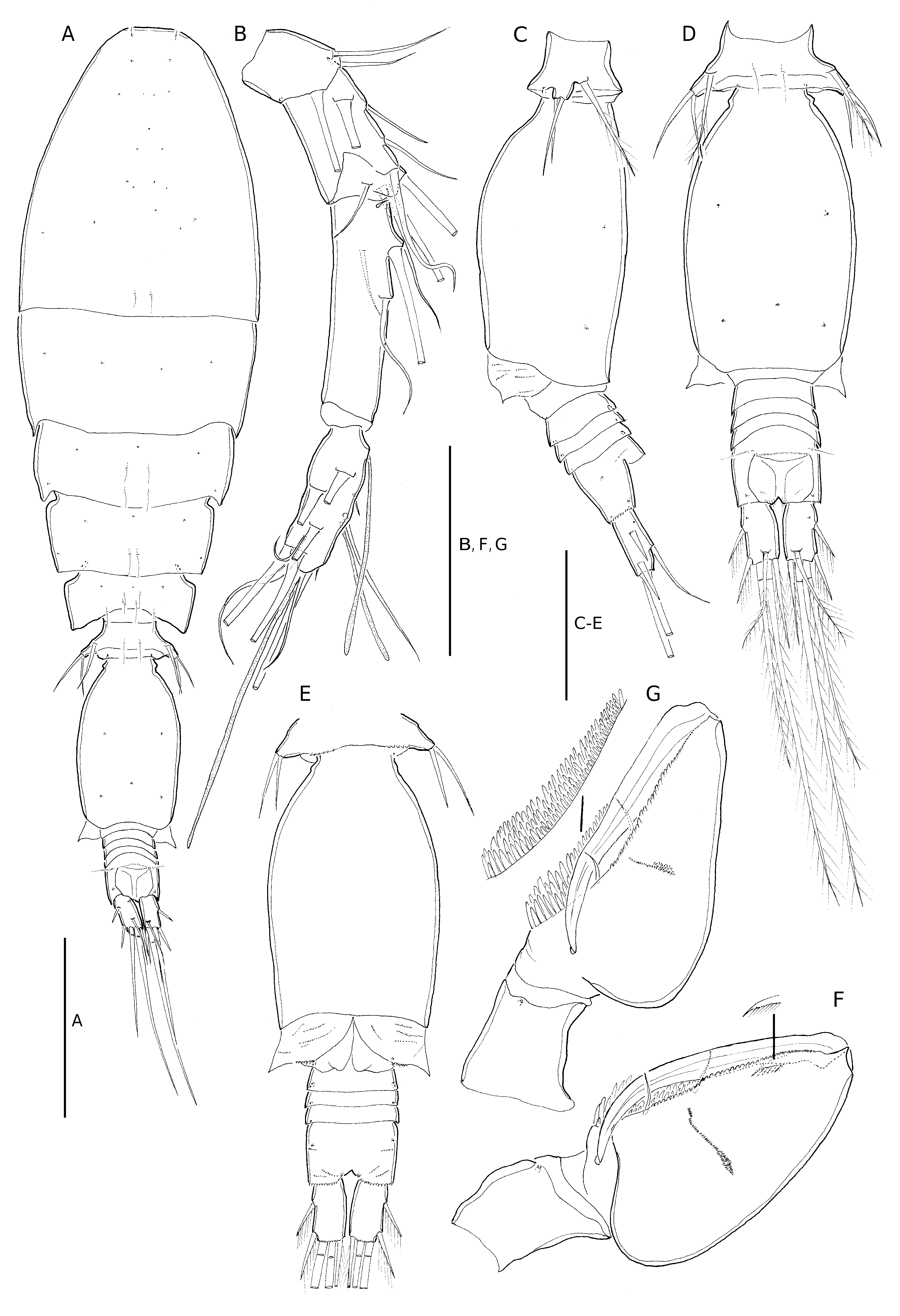
( Figs 6-9 View FIG View FIG View FIG View FIG ; 13B, C View FIG ; Tables 2-4)
urn:lsid:zoobank.org:act:
TYPE LOCALITY. — Northeastern equatorial Pacific Ocean ( 10°30’N, 131°20’W, 0-100 m).
TYPE MATERIAL. — Holotype. 1♀; NIBRIV0000838008 ; 10°30’N, 131°20’W; 0-100 m; dissected and mounted on 10 slides, collected from the type locality on 21.VIII.2009 by D. J. Ham. GoogleMaps
Paratypes. 3 ♀; NIBRIV0000838009-011; 10°30’N, 131°20’W; 0-100 m; each dissected and mounted on 9 or 10 slides, respectively (lost a slide of urosome with P5 of the third paratype). — 3 ♂; NIBRIV0000838012-014; 10°30’N, 131°20’W; 0-100 m; each dissected and mounted on 10 slides, respectively. — 4♀, 3♂ kept in one vial in alcohol, MNHN-IU-2019-2283. All specimens are from the type locality.
ETYMOLOGY. — The species is named after research vehicle ‘Onnuri’ of the Korea Institute of Ocean Science and Technology (KIOST) to recognize contributions to research activities in the northeastern equatorial Pacific Ocean.
DESCRIPTION
Female
Body length. 784-823 Μm, based on four specimens.
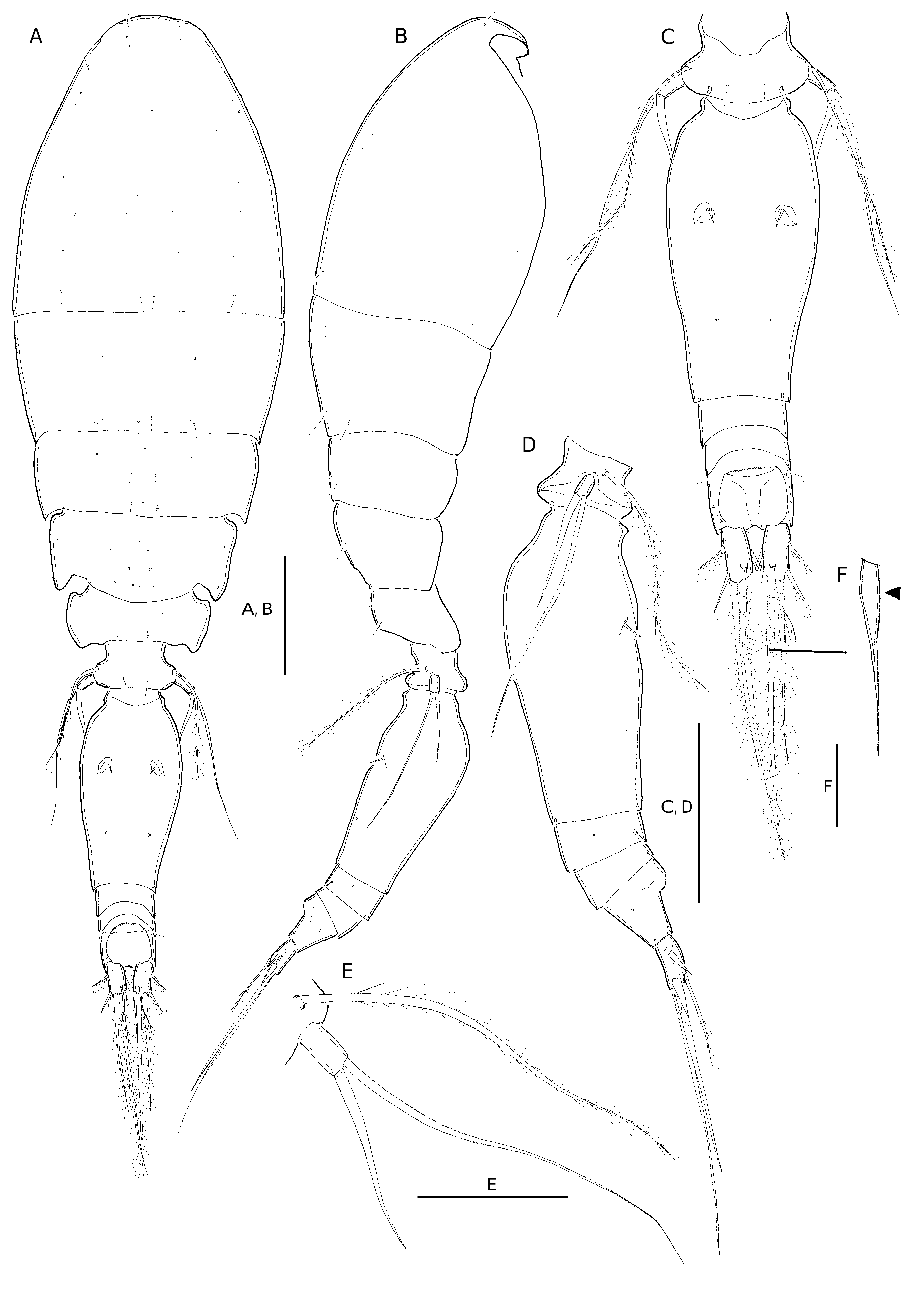
Exoskeleton moderately chitinized. Prosome about 1.9 times length of urosome, excluding caudal rami 1.7 times urosome length including caudal rami. P2-bearing somite without conspicuous dorsoposterior projection in lateral aspect ( Fig. 6B View FIG ). Integumental pores on prosome as indicated in Fig. 6A View FIG . Pleural areas of P4-bearing somite with rounded posterolateral corners. One pair of secretory pores discernible on first postgenital somite ( Fig. 6D View FIG ).
Genital double-somite. 2.0 times as long as maximum width (measured in dorsal aspect) and about 2.2 times as long as postgenital somites combined; largest width measured at anterior one third; posterior part tapering gradually ( Fig. 6C View FIG ); surface covered with numerous minute pores or pits near genital apertures (inset of Fig. 13B View FIG ). Paired genital apertures located dorsally at about 1/3 of distance from anterior margin of genital double-somite ( Fig. 6C View FIG ); armature represented by one long spine and minute spinule ( Fig. 13B View FIG ). Secretory pores on dorsal surface as indicated in Fig. 6C View FIG .
Anal somite. About 1.4 times wider than long; slightly longer than caudal rami ( Fig. 6C View FIG ). Ornamentation as in T. komo n. sp.
Caudal ramus. About two times as long as wide. Seta VII about half length of seta IV; seta VI almost same length as seta VII, swollen at base (arrowed in Fig. 6F View FIG ).
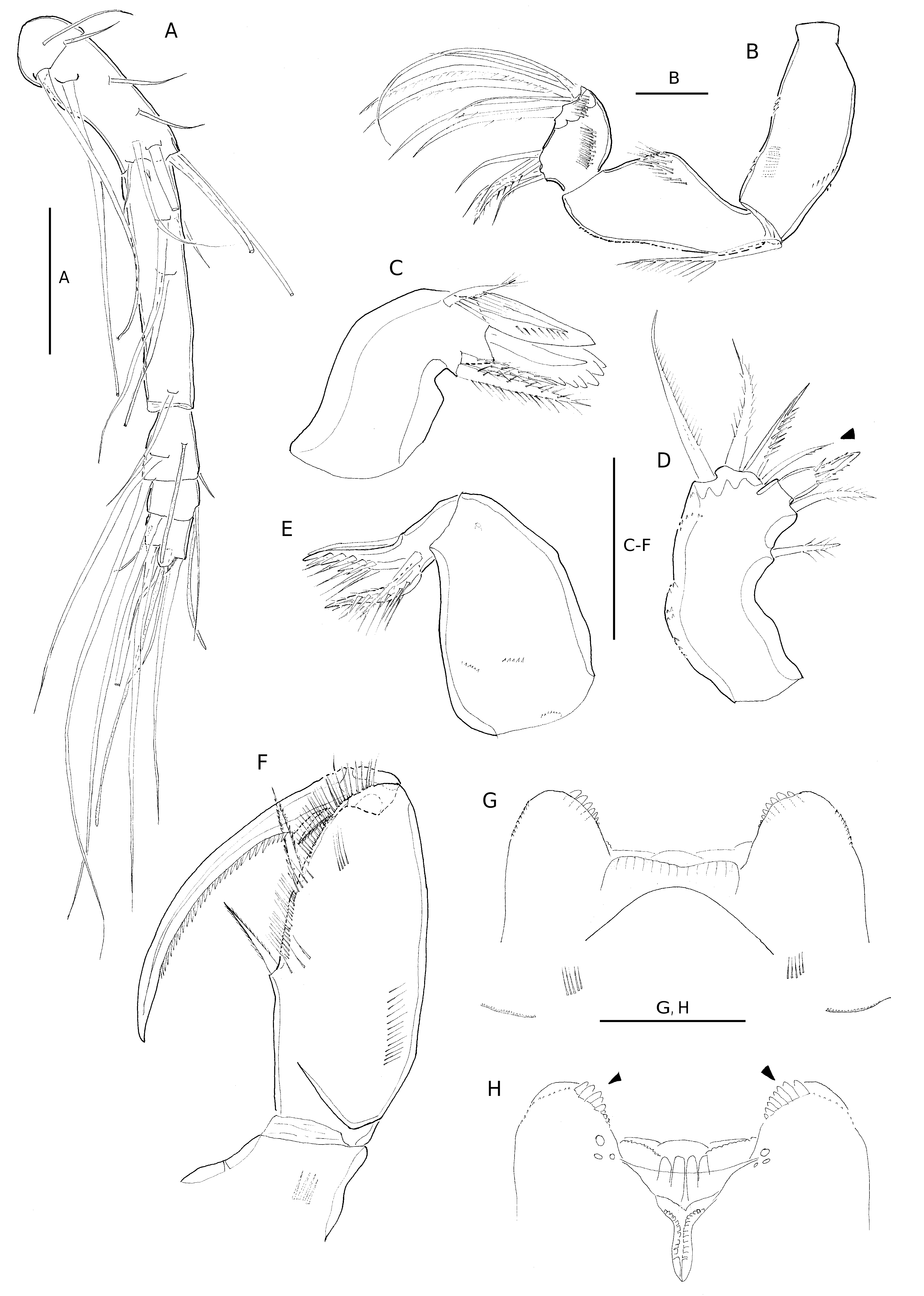
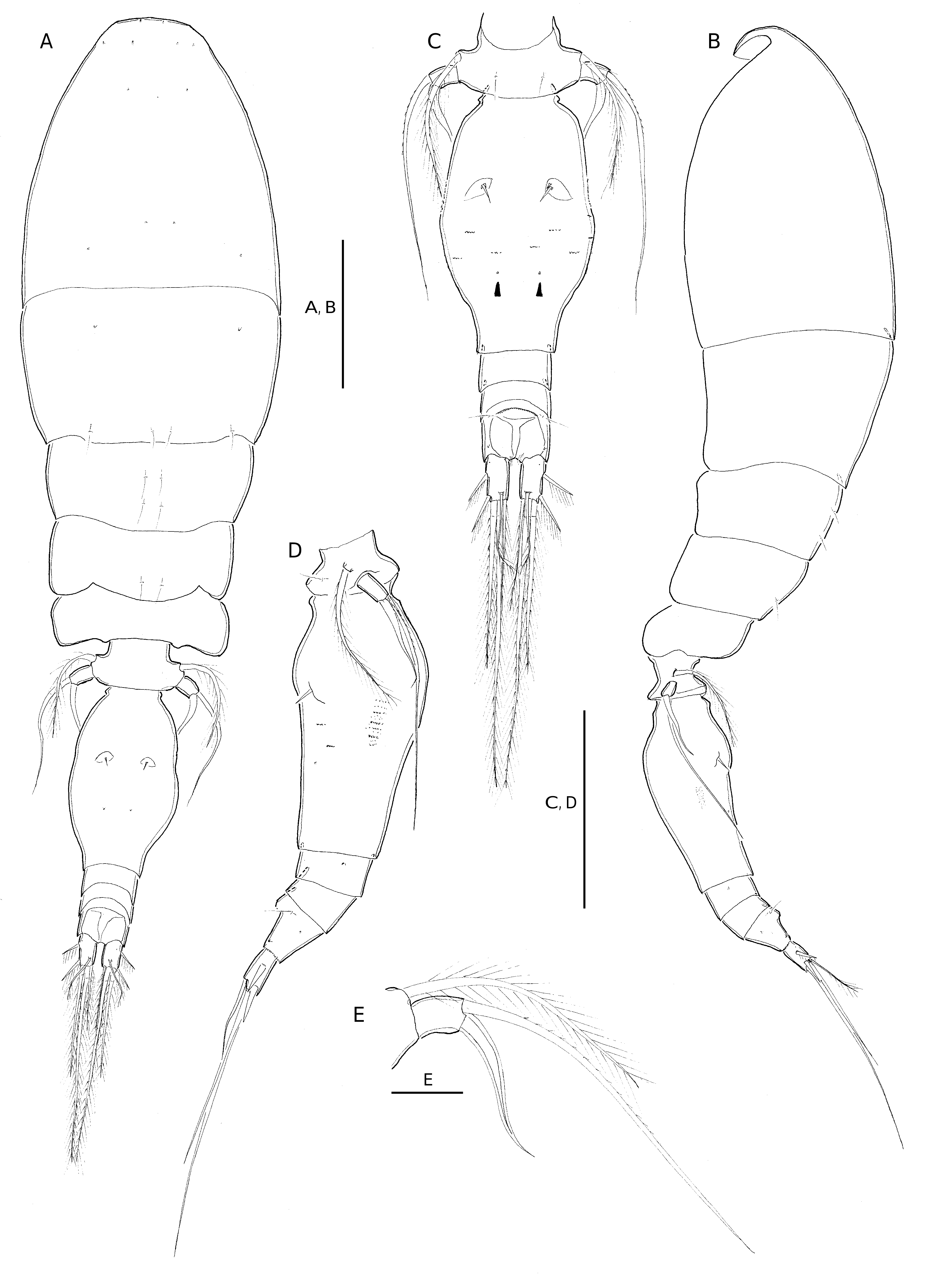
Antennule ( Fig. 7A View FIG ). 6-segmented. Armature formula as for T. komo n. sp.
Antenna ( Fig. 7B View FIG ). 3-segmented. Distal endopod segment with armature and ornamentation as in T. komo n. sp., except for seta I of lateral armature sparsely pinnate, and seta III bipinnate at distal part.
Labrum ( Fig. 7G, H View FIG ). Similar to T. komo n. sp., except for each lobe with stronger and fewer dentiform processes than in T. komo n. sp.
Mandible ( Fig. 7C View FIG ). Maxillule ( Fig. 7D View FIG ) and maxilla ( Fig. 7E View FIG ) as for T. komo n. sp., except for innermost element on outer lobe of maxillule ornamented with small spinules (arrowed in Fig. 7D View FIG ).
Maxilliped ( Fig. 7F View FIG ). Similar to T. komo n. sp., basis with two bipinnate spiniform elements, nearly equal in length, distal one slightly longer. Distal endopod segment (claw) with row of strong pinnules along proximal 4/5 of concave margin.
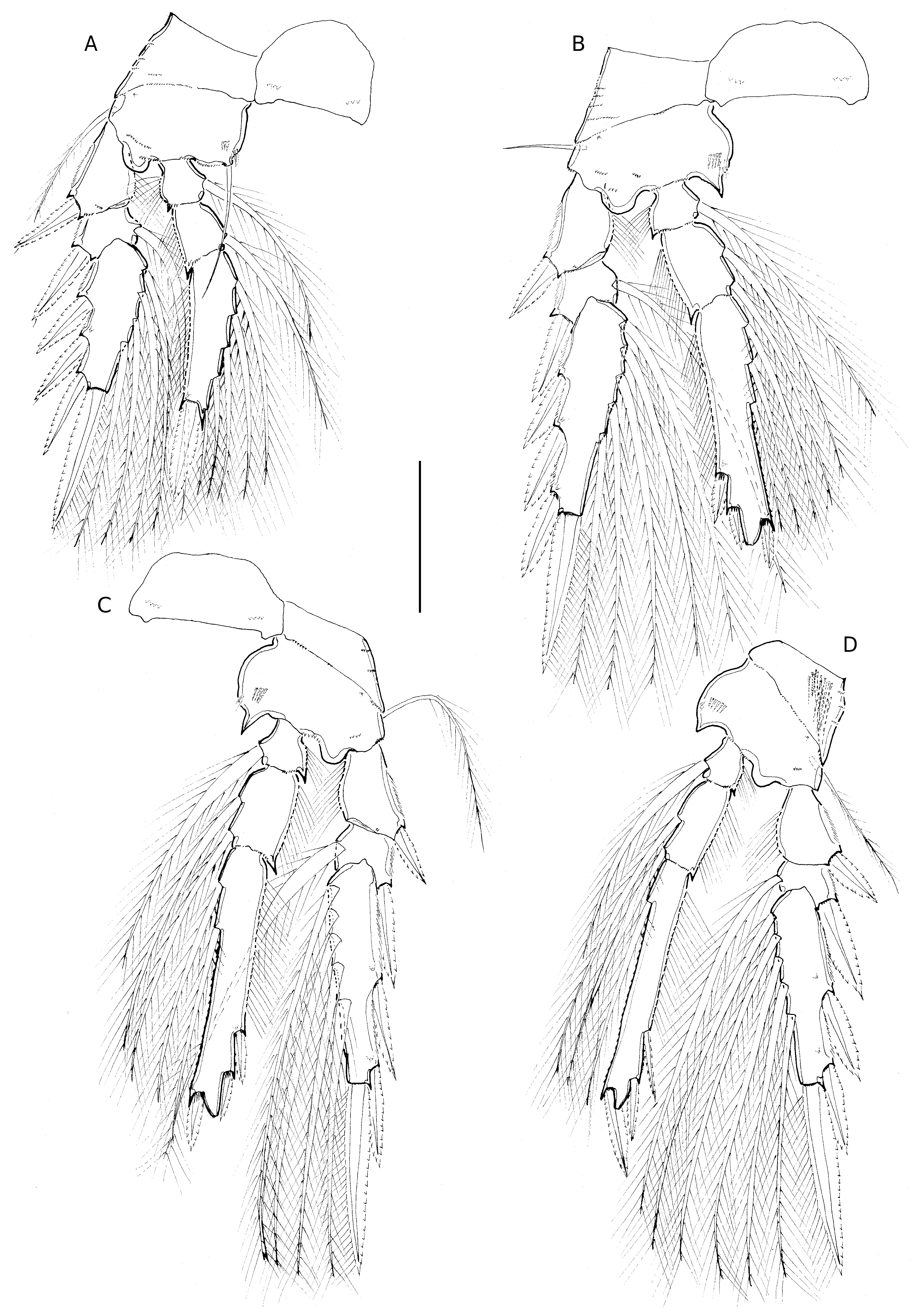
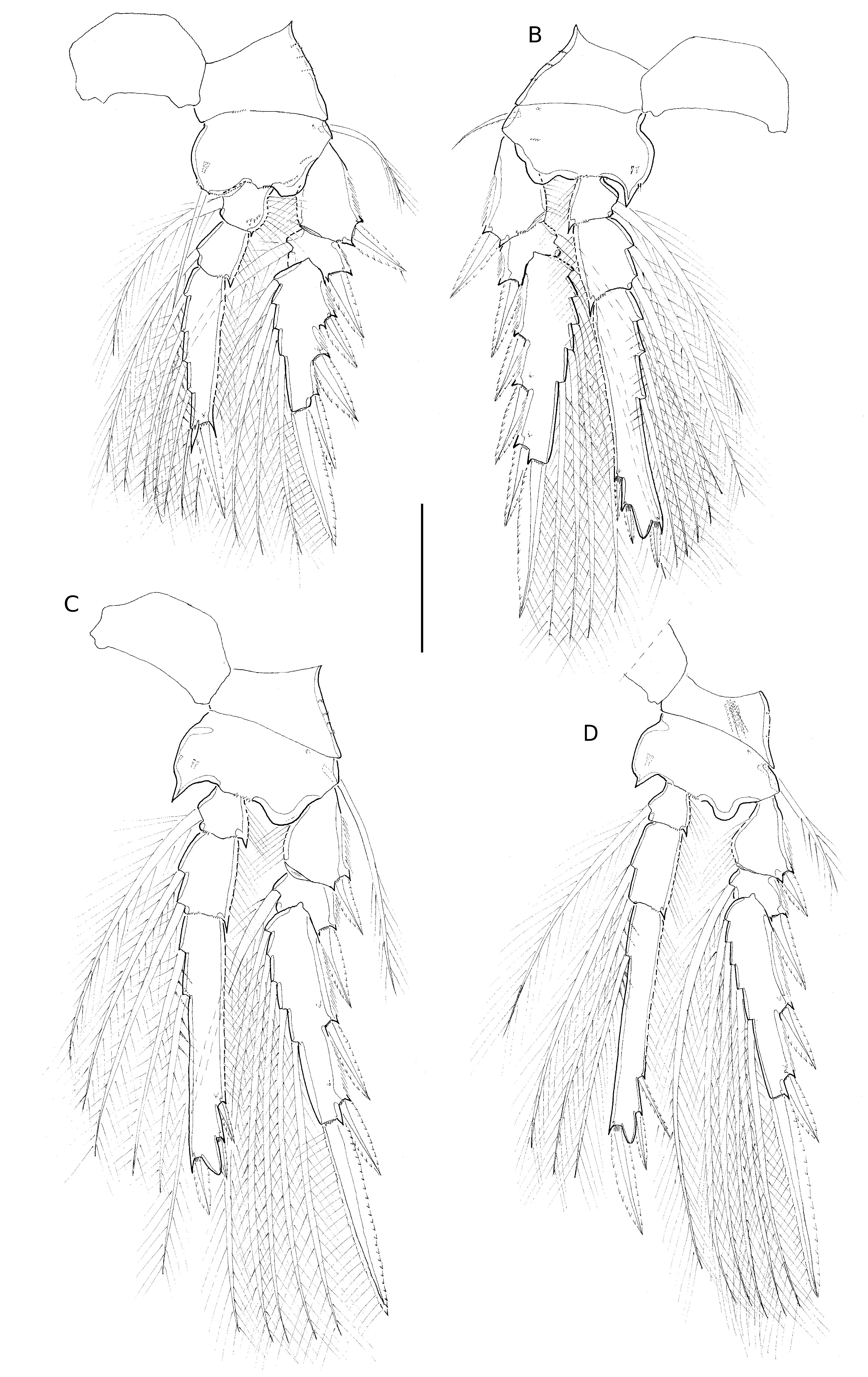
Swimming legs 1-4. Biramous ( Fig. 8 View FIG A-D). With armature as in T. komo n. sp. Intercoxal sclerites of P1-P3 ornamented with few spinules on posterior face ( Fig. 8A, B, C View FIG ); intercoxal sclerite of P4 not observed. Coxae and bases of P1-P4 with ornamentation as shown in Fig. 8 View FIG A-D. Basis of P4 with outer seta shorter than in T. komo n. sp. ( Fig. 8D View FIG ).
Exopods. Similar to T. komo n. sp., except for length of outer spine on P3 exp-1 shorter than in T.komo n. sp. ( Fig. 8C View FIG ). Length ratio of outer spine on exp-1 relative to outer spine on exp-2 of P3 and P4 somewhat smaller than in T. komo n. sp. ( Table 3).
Endopods. Distal margin of P2-P4 produced into conical process with apical pore. Length ratios of spines different from T. komo n. sp. with length data of spines of five specimens as shown in Table 2; length ranges of outer subdistal spine (OSDS) and outer distal spine (ODS) relative to distal spine (DS) given in Table 3.
P5 ( Fig. 6E View FIG ). With outer basal seta very long and plumose at distal part; exopod segment free. Exopod about 1.9 times longer than wide, bearing two naked setae, bearing stout inner seta and extremely long, slender outer seta about twice of length of inner seta, reaching 4/5 the length of genital double-somite from anterior margin, as far as secretory pores on dorsal surface.
P6 ( Fig. 6F View FIG ). Represented by operculum closing off each genital aperture, ornamented with long spine and minute spinule ( Fig. 13B View FIG ).
Male
Body length. 570-604 Μm, based on three specimens. Sexual dimorphism in antennule, maxilliped, P5-P6, caudal ramus, and in genital segmentation. Prosome 1.8 times urosome length, including caudal rami ( Fig. 9A View FIG ). Cephalosome and P1 bearing somite with conspicuous lateral patterns of pore patches ( Figs 9B View FIG ; 13C View FIG ). Posterior margin of P5-bearing somite with paired row of minute denticles or spinules ventrally ( Fig. 9E View FIG ).
Genital somite ( Fig. 9D View FIG ). About 1.5 times longer than wide.
Caudal rami. About 1.4 times longer than wide, shorter than in female. Caudal seta with proportional lengths as in female, seta VI swollen at base as in female (arrowed in Fig. 9I View FIG ). Dorsal surface of genital somite with three secretory pores as indicated in Fig. 9D View FIG . Surface of genital flaps ornamented with several rows of small spinules ( Fig. 9E, F View FIG ).
Antennule ( Fig. 9C View FIG ). 4-segmented; armature formula as for T. komo n. sp.
Maxilliped ( Fig. 9G, H View FIG ). 3-segmented. Surface ornamentation on syncoxa not discernible, except for single secretory pore at inner distal margin. Basis robust, with small naked setae within longitudinal cleft, proximal seta about same length as distal one; anterior surface with one-two transverse spinular rows and row of small flat spinules along inner margin; posterior surface with rows of short spinules of graduated length along palmar margin.
Swimming legs. With armature and ornamentation as in female, length data of endopodal spines of three males as shown in Table 2; length ranges of outer subdistal spine ( OSDS) and outer distal spine ( ODS) relative to distal spine given in Table 4; generally similar to females (cf. Table 3).
P5 ( Fig. 9D, F View FIG ). Exopod not delimited from somite, shorter than in female; outer exopodal seta unornamented and almost equal in length to inner seta; outer basal seta naked and much shorter than in female.
P6. Represented by posterolateral flap closing off genital aperture on either side; covered by pattern of spinules as shown in Fig. 9E View FIG ; posterolateral corners protruding laterally and visible in dorsal aspect ( Fig. 9A, D View FIG ).
REMARKS
Among species of the similis -subgroup of Triconia , T. onnuri n. sp. is closely related to T. similis , based on the body size and the form of the genital double-somite in the female, but differs in the relative lengths of the outer exopodal seta and the outer basal seta on P5, with both setae reaching over half the distance from anterior to posterior margin of the genital double-somite, and the form of caudal seta VI, which is swollen at its base. The combination of these characters separates the new species also from other described species of the similis -subgroup. Furthermore, T. onnuri n. sp. can be identified by the length to width ratio of P5 exopod, which is intermediate (1.9:1) between those of other species of the similis -subgroup (most are less than 1.5:1, except 3: 1 in T. recta ); and in some minor differences in the proportional spine lengths on the endo- and /or exopods of P2-P4. Especially on the endopod of P4, the length ratio values of outer subdistal spine ( OSDS) relative to distal spine (DS) is higher in T. onnuri n. sp. ( OSDS:DS=0.73-0.86:1) than the range of single values reported for T. similis from three studies ( OSDS:DS=0.62-0.67:1, Table 3).
The male of T. onnuri n. sp. shows a distinct modification of seta VI on the caudal ramus, which is basally swollen ( Fig. 8I View FIG ) as in the female ( Fig. 5F View FIG ). Modifications in the form of caudal setae have not been observed in other species of Triconia so far, but have been reported for other oncaeid species, such as species of Spinoncaea (Böttger-Schnack 2003) and Epicalymma (Böttger-Schnack 2009) , where they occur in both sexes as well. Thus, in T. onnuri n. sp., the modification of the caudal setae can be used as an additional tool for separating the species from closely related species within the genus. Also in the male, the proportional spine lengths on the endopod of P2-P4 and the length range of the exopodal spines on exp-1 of P3-P4 are similar to the female.
Triconia denticula Wi, Shin & Soh, 2011
( Figs 10-12 View FIG View FIG View FIG ; Tables 2, 3)
Triconia denticula Wi, Shin & Soh, 2011: 590-595 , figs 2-4, 9A, B, F ( ♀).
TYPE LOCALITY. — East China Sea (south of Cheju Island)
MATERIAL EXAMINED. — East China Sea. 3 ♀; NIBRIV-0000838015-017; each dissected and mounted on 10 slides, respectively; all specimens collected from the northeastern equatorial Pacific Ocean; station BN09-02-01; 10°30’N, 131°20’W, 0-100 m; 21. VII.2009 by D. J. Ham.
DESCRIPTION
Female
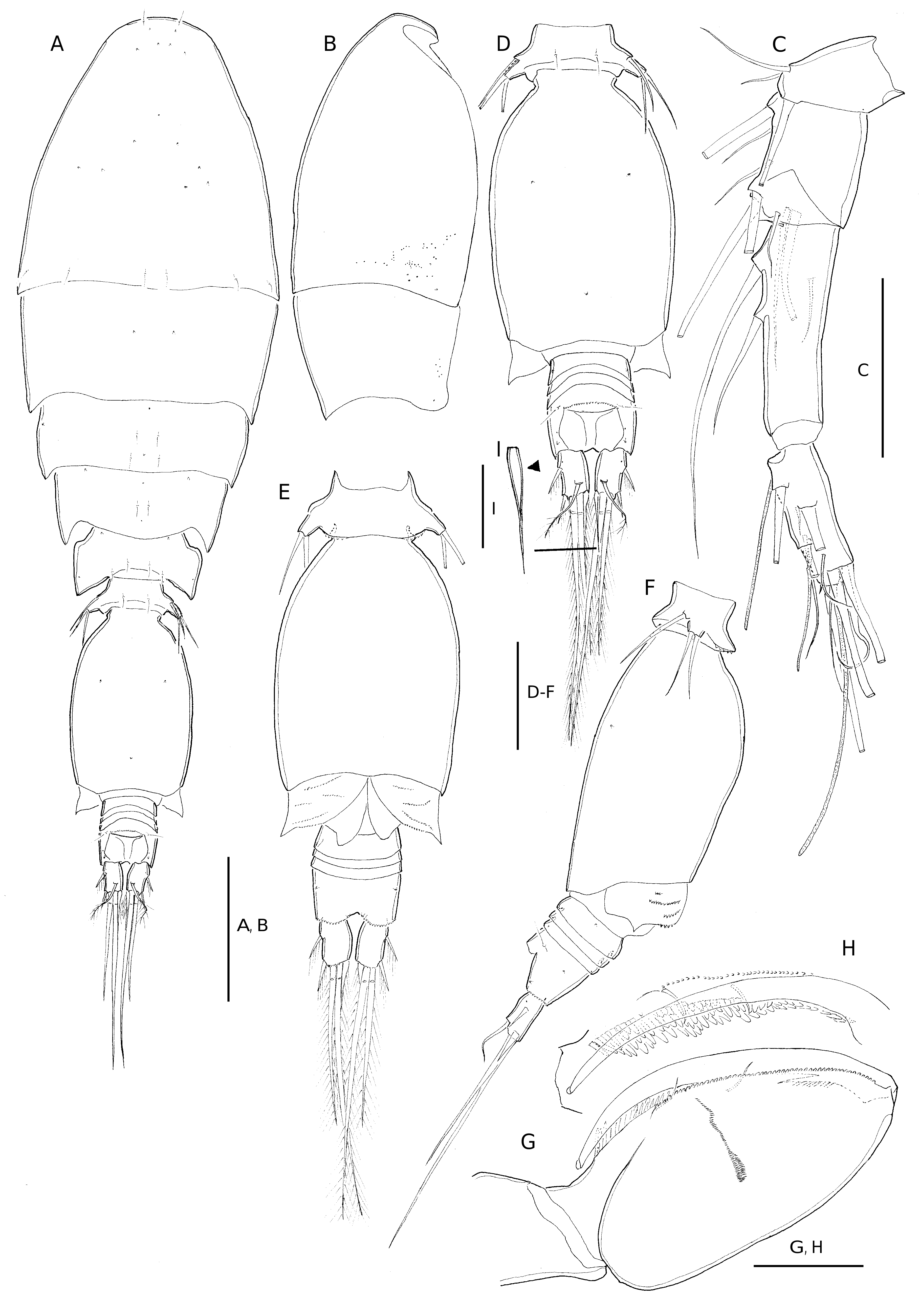
Body length. 648-663 Μm, based on three specimens. Exoskeleton moderately chitinized. Prosome about 2.1 times length of urosome, excluding caudal rami 1.9 times urosome length including caudal rami.P2-bearing somite without conspicuous dorsoposterior projection in lateral aspect ( Fig. 10B View FIG ). Integumental pores on prosome as indicated in Fig. 10A View FIG . Pleural areas of P4-bearing somite with rounded posterolateral corners. One pair of secretory pores discernible on first postgenital somite ( Fig. 10D View FIG ).
Genital double-somite. 1.7 times as long as maximum width (measured in dorsal aspect) and about 2.1 times as long as postgenital somites combined; largest width measured just posterior to mid-level; lateral margins of genital double-somite sinuous anterior to level of maximum width, tapering posteriorly ( Fig. 10C View FIG ); dorsal and lateral surface ornamented with several rows of spinules ( Fig. 10C, D View FIG ); anterior part of genital double-somite protruding dorsally ( Fig. 10D View FIG ). Paired genital apertures located at about 2/5 of distance along genital doublesomite from anterior margin. Paired secretory pores on dorsal surface located at about 2/3 of distance along double-somite from anterior margin (arrowed in Fig. 10C View FIG ).
Anal somite. Slightly wider than long; slightly longer than caudal rami ( Fig. 10C View FIG ). Ornamentation as in T. komo n. sp. ( Fig. 10C, D View FIG ).
Caudal ramus. About 1.6 times as long as wide; with seta VII slightly longer than half the length of seta IV and shorter than seta VI ( Fig. 10C View FIG ).
Antennule. 6-segmented, armature formula as for T. komo n. sp. ( Fig. 11A View FIG ).
Antenna ( Fig. 11B View FIG ). 3-segmented. Distal endopod segment with distal armature consisting of five curved setae, long naked (E), entire unipinnate (A), sparsely pinnate (B-D), setae A-D of graduated length with seta D shortest, and two slender bare setae (F+G), seta G shortest.
Labrum ( Fig. 11G, H View FIG ). With medial concavity between lobes covered anteriorly by single long hyaline lamella, lobes with dentiform processes around outer ventral margin more slender than in T. komo n. sp. Anterior surface with paired row of long setules and free margin of integumental pockets either side of median swelling ornamented with minute denticles. Posterior wall of medial concavity ornamented with four long sclerotized “teeth”, posterior surface with group of four secretory pores located on proximal part of each lobe ( Fig. 11H View FIG ).
Mandible ( Fig. 11C View FIG ). Maxillule ( Fig. 11D View FIG ) and maxilla ( Fig. 11E View FIG ) as for T. komo n. sp., with slight differences in surface ornamentation of coxa on mandible, and on syncoxa on maxilla.
Maxilliped ( Fig. 11F View FIG ). With ornamentation of syncoxa as figured. Basis with two bipinnate spiniform elements, nearly equal in length; fringe of long pinnules between distal seta and articulation with endopod, row of long spinules between proximal and distal setae; short transverse row of long setules near distal seta on anterior surface and additional longitudinal row near outer margin. Distal endopod segment (claw) with row of pinnules along proximal 2/3 of concave margin. Other elements as in T. komo n. sp.
Swimming legs 1-4 biramous ( Fig. 12 View FIG A-D). With armature and ornamentation as in T. komo n. sp. Intercoxal sclerites well developed, without ornamentation. Bases with short (P1, P2, P4) or very long (P3) outer seta.
Exopods. With ornamentation similar to T. komo n. sp. Distal spine almost equal in length to (P4) or shorter (P1, P2, P3) than distal exopod segment. Length of exopodal spine on exp-1 and exp-2 of P3 and P4 shorter than in T. komo n. sp. ( Fig. 12C, D View FIG ). Length ratio of outer spine on exp-1 relative to outer spine on exp-2 of P3 and P4 similar to T. komo n. sp. ( Table 3).
Endopods. Distal margin of P2-P4 produced into conical process ( Fig. 12 View FIG B-D), process with apical pore. Length data of spines on P2-P4 enp-3 of three specimens as shown in Table 2; length ranges of outer subdistal spine ( OSDS) and outer distal spine ( ODS) relative to distal spine (DS) given in Table 3.
P5 ( Fig. 10E View FIG ). Comprising long plumose outer basal seta and free unornamented exopod segment. Exopod longer than wide, bearing long slender seta (outer seta) ornamented with fine spinules along outer margin and a stout curved seta (inner seta), swollen at base and naked; outer seta about twice longer than inner seta.
P6 ( Fig. 10C View FIG ). Represented by operculum closing off each genital aperture, ornamented with spine and minute spinule (visible under light microscope).
Male.
Unknown
REMARKS
The original description of T. denticula by Wi et al. (2011) was based on female specimens from the south of Cheju Island of Korea (the East China Sea). As mentioned by Wi et al. (2011), this species closely resembles T. rufa in the form of the genital double-somite and the relative length of the outer basal seta and exopodal setae on P5, but differs: 1) in the absence of the dorsoposterior projection on the P2-bearing somite, which is present in T. rufa , a species of the conifera -subgroup; and 2) in the length to width ratio of P5 exopod, being 1.5 times longer than wide, whereas the length is 2.8 times longer than wide in T. rufa (Böttger-Schnack 1999) .
Specimens of T. denticula from the northeastern equatorial Pacific Ocean agree in almost all morphological characters with the original description of the species from Korean waters, based on the figures published by Wi et al. (2011). But they exhibited some variability in the length ratio of the prosome to urosome (including CR), with the Pacific specimens being larger (1.9:1) than those in the Korean waters (1.7:1); in the length to width ratio of the genital double-somite, being somewhat more elongate (1.7:1) than in the Korean waters specimens (1.5:1); in the length ratio of the caudal ramus to anal somite, which is shorter (0.6:1) in the Pacific specimens, compared to those in the Korean waters (about same length); and in body size, being somewhat smaller (648-663 Μm) in our study area than in the Korean specimens (660-710 Μm) (cf. Table 3).
Wi et al. (2011) mentioned that the exopodal spines on the first exopod segment in P3 and P4 of T. denticula are shortest among the species examined. However, in that study the definition of the proportional lengths of the exopodal spines is not sufficiently clear, as it can be misinterpreted where e.g. “[…] the tip of exopodal spine is reaching […]”. In order to provide more precise information, we calculated the length of the spine on exp-1 relative to the spine on exp-2 of P3 and P 4 in T. denticula from the Korean waters ( paratype) in NIBR and from the Pacific Ocean. We also measured the lengths of the endopodal spines on P2-P 4 in specimens of the two areas. The resulting length ratios are given in Table 3. The recalculated ratio values for Korean specimens are largely overlapping with the range of values reported for specimens from the equatorial Pacific Ocean; only the ratio values for spines on P2 enp-3 are not completely overlapping and are tentatively higher for Korean specimens than for the open Pacific specimens.
T. denticula from the Korean waters described by Wi et al. (2011) exhibited numerous small scales on the surfaces of the genital double-somite, the anal somite and the caudal ramus, which were discerned by a using scanning electron microscope. Actually, these structures are hardly discernible with a light microscope due to their small size. In the Pacific specimens, the surface ornamentation of the genital double-somite was probably not fully discerned, but just on the dorsolateral part at half length of the genital double-somite. Also, T. denticula specimens from the two areas seemed to differ in the position of paired integumental pores on the dorsal surface of the genital double-somite, which are located at approximately 2/3 of distance along genital double-somite from anterior margin in the Pacific specimen and in the same vertical line with the genital apertures (cf. Fig. 9A, C View FIG ), while in the specimen from the Korean waters they were figured as being situated at about half the distance along the genital double-somite from anterior margin and much closer to the lateral margin of the double-somite ( Wi et al. 2011: fig. 3C). However, upon reexamination of the undissected female paratype of T. denticula (NIBRIV0000214678) during the present study, it was found that the position of paired integumental pores was similar to their position in the Pacific specimens.
The type material of T. denticula from the Korean waters which was loaned from the collections of NIBR and reexamined by the senior author of the present study turned out to be insufficient for taxonomic comparisons due to the following reasons:
1) The holotype sample (NIBRIV0000214676) nominated as: “ Holotype female dissected and mounted on 1 glass slide” ( Wi et al. 2011: 590) was found to contain an undissected female specimen of the dentipes -subgroup of Triconia on this slide.
2) The first paratype sample (NIBRIV0000214677) nominated as: “ 2 females dissected and mounted on 3 slides”, was in very poor condition and it was difficult to make out the contents on the slides. Recognizable contents were present in only one of the three slides.
3) The second paratype sample (NIBRIV0000214678) nominated as: “[…] 2 undissected females in 1 vial […]” ( Wi et al. 2011: 590) included one female of T. denticula and one female of a species of the dentipes -subgroup of Triconia .
In conclusion, a fundamental revision of the type material of T. denticula is required.
Triconia denticula Wi, Shin & Soh, 2011
( Figs 10-12 View FIG View FIG View FIG ; Tables 2, 3)
Triconia denticula Wi, Shin & Soh, 2011: 590-595 , figs 2-4, 9A, B, F ( ♀).
TYPE LOCALITY. — East China Sea (south of Cheju Island)
MATERIAL EXAMINED. — East China Sea. 3 ♀; NIBRIV-0000838015-017; each dissected and mounted on 10 slides, respectively; all specimens collected from the northeastern equatorial Pacific Ocean; station BN09-02-01; 10°30’N, 131°20’W, 0-100 m; 21. VII.2009 by D. J. Ham.
DESCRIPTION
Female
Body length. 648-663 Μm, based on three specimens. Exoskeleton moderately chitinized. Prosome about 2.1 times length of urosome, excluding caudal rami 1.9 times urosome length including caudal rami.P2-bearing somite without conspicuous dorsoposterior projection in lateral aspect ( Fig. 10B View FIG ). Integumental pores on prosome as indicated in Fig. 10A View FIG . Pleural areas of P4-bearing somite with rounded posterolateral corners. One pair of secretory pores discernible on first postgenital somite ( Fig. 10D View FIG ).
Genital double-somite. 1.7 times as long as maximum width (measured in dorsal aspect) and about 2.1 times as long as postgenital somites combined; largest width measured just posterior to mid-level; lateral margins of genital double-somite sinuous anterior to level of maximum width, tapering posteriorly ( Fig. 10C View FIG ); dorsal and lateral surface ornamented with several rows of spinules ( Fig. 10C, D View FIG ); anterior part of genital double-somite protruding dorsally ( Fig. 10D View FIG ). Paired genital apertures located at about 2/5 of distance along genital doublesomite from anterior margin. Paired secretory pores on dorsal surface located at about 2/3 of distance along double-somite from anterior margin (arrowed in Fig. 10C View FIG ).
Anal somite. Slightly wider than long; slightly longer than caudal rami ( Fig. 10C View FIG ). Ornamentation as in T. komo n. sp. ( Fig. 10C, D View FIG ).
Caudal ramus. About 1.6 times as long as wide; with seta VII slightly longer than half the length of seta IV and shorter than seta VI ( Fig. 10C View FIG ).
Antennule. 6-segmented, armature formula as for T. komo n. sp. ( Fig. 11A View FIG ).
Antenna ( Fig. 11B View FIG ). 3-segmented. Distal endopod segment with distal armature consisting of five curved setae, long naked (E), entire unipinnate (A), sparsely pinnate (B-D), setae A-D of graduated length with seta D shortest, and two slender bare setae (F+G), seta G shortest.
Labrum ( Fig. 11G, H View FIG ). With medial concavity between lobes covered anteriorly by single long hyaline lamella, lobes with dentiform processes around outer ventral margin more slender than in T. komo n. sp. Anterior surface with paired row of long setules and free margin of integumental pockets either side of median swelling ornamented with minute denticles. Posterior wall of medial concavity ornamented with four long sclerotized “teeth”, posterior surface with group of four secretory pores located on proximal part of each lobe ( Fig. 11H View FIG ).
Mandible ( Fig. 11C View FIG ). Maxillule ( Fig. 11D View FIG ) and maxilla ( Fig. 11E View FIG ) as for T. komo n. sp., with slight differences in surface ornamentation of coxa on mandible, and on syncoxa on maxilla.
Maxilliped ( Fig. 11F View FIG ). With ornamentation of syncoxa as figured. Basis with two bipinnate spiniform elements, nearly equal in length; fringe of long pinnules between distal seta and articulation with endopod, row of long spinules between proximal and distal setae; short transverse row of long setules near distal seta on anterior surface and additional longitudinal row near outer margin. Distal endopod segment (claw) with row of pinnules along proximal 2/3 of concave margin. Other elements as in T. komo n. sp.
Swimming legs 1-4 biramous ( Fig. 12 View FIG A-D). With armature and ornamentation as in T. komo n. sp. Intercoxal sclerites well developed, without ornamentation. Bases with short (P1, P2, P4) or very long (P3) outer seta.
Exopods. With ornamentation similar to T. komo n. sp. Distal spine almost equal in length to (P4) or shorter (P1, P2, P3) than distal exopod segment. Length of exopodal spine on exp-1 and exp-2 of P3 and P4 shorter than in T. komo n. sp. ( Fig. 12C, D View FIG ). Length ratio of outer spine on exp-1 relative to outer spine on exp-2 of P3 and P4 similar to T. komo n. sp. ( Table 3).
Endopods. Distal margin of P2-P4 produced into conical process ( Fig. 12 View FIG B-D), process with apical pore. Length data of spines on P2-P4 enp-3 of three specimens as shown in Table 2; length ranges of outer subdistal spine ( OSDS) and outer distal spine ( ODS) relative to distal spine (DS) given in Table 3.
P5 ( Fig. 10E View FIG ). Comprising long plumose outer basal seta and free unornamented exopod segment. Exopod longer than wide, bearing long slender seta (outer seta) ornamented with fine spinules along outer margin and a stout curved seta (inner seta), swollen at base and naked; outer seta about twice longer than inner seta.
P6 ( Fig. 10C View FIG ). Represented by operculum closing off each genital aperture, ornamented with spine and minute spinule (visible under light microscope).
Male.
Unknown
REMARKS
The original description of T. denticula by Wi et al. (2011) was based on female specimens from the south of Cheju Island of Korea (the East China Sea). As mentioned by Wi et al. (2011), this species closely resembles T. rufa in the form of the genital double-somite and the relative length of the outer basal seta and exopodal setae on P5, but differs: 1) in the absence of the dorsoposterior projection on the P2-bearing somite, which is present in T. rufa , a species of the conifera -subgroup; and 2) in the length to width ratio of P5 exopod, being 1.5 times longer than wide, whereas the length is 2.8 times longer than wide in T. rufa (Böttger-Schnack 1999) .
Specimens of T. denticula from the northeastern equatorial Pacific Ocean agree in almost all morphological characters with the original description of the species from Korean waters, based on the figures published by Wi et al. (2011). But they exhibited some variability in the length ratio of the prosome to urosome (including CR), with the Pacific specimens being larger (1.9:1) than those in the Korean waters (1.7:1); in the length to width ratio of the genital double-somite, being somewhat more elongate (1.7:1) than in the Korean waters specimens (1.5:1); in the length ratio of the caudal ramus to anal somite, which is shorter (0.6:1) in the Pacific specimens, compared to those in the Korean waters (about same length); and in body size, being somewhat smaller (648-663 Μm) in our study area than in the Korean specimens (660-710 Μm) (cf. Table 3).
Wi et al. (2011) mentioned that the exopodal spines on the first exopod segment in P3 and P4 of T. denticula are shortest among the species examined. However, in that study the definition of the proportional lengths of the exopodal spines is not sufficiently clear, as it can be misinterpreted where e.g. “[…] the tip of exopodal spine is reaching […]”. In order to provide more precise information, we calculated the length of the spine on exp-1 relative to the spine on exp-2 of P3 and P 4 in T. denticula from the Korean waters ( paratype) in NIBR and from the Pacific Ocean. We also measured the lengths of the endopodal spines on P2-P 4 in specimens of the two areas. The resulting length ratios are given in Table 3. The recalculated ratio values for Korean specimens are largely overlapping with the range of values reported for specimens from the equatorial Pacific Ocean; only the ratio values for spines on P2 enp-3 are not completely overlapping and are tentatively higher for Korean specimens than for the open Pacific specimens.
T. denticula from the Korean waters described by Wi et al. (2011) exhibited numerous small scales on the surfaces of the genital double-somite, the anal somite and the caudal ramus, which were discerned by a using scanning electron microscope. Actually, these structures are hardly discernible with a light microscope due to their small size. In the Pacific specimens, the surface ornamentation of the genital double-somite was probably not fully discerned, but just on the dorsolateral part at half length of the genital double-somite. Also, T. denticula specimens from the two areas seemed to differ in the position of paired integumental pores on the dorsal surface of the genital double-somite, which are located at approximately 2/3 of distance along genital double-somite from anterior margin in the Pacific specimen and in the same vertical line with the genital apertures (cf. Fig. 9A, C View FIG ), while in the specimen from the Korean waters they were figured as being situated at about half the distance along the genital double-somite from anterior margin and much closer to the lateral margin of the double-somite ( Wi et al. 2011: fig. 3C). However, upon reexamination of the undissected female paratype of T. denticula (NIBRIV0000214678) during the present study, it was found that the position of paired integumental pores was similar to their position in the Pacific specimens.
The type material of T. denticula from the Korean waters which was loaned from the collections of NIBR and reexamined by the senior author of the present study turned out to be insufficient for taxonomic comparisons due to the following reasons:
1) The holotype sample (NIBRIV0000214676) nominated as: “ Holotype female dissected and mounted on 1 glass slide” ( Wi et al. 2011: 590) was found to contain an undissected female specimen of the dentipes -subgroup of Triconia on this slide.
2) The first paratype sample (NIBRIV0000214677) nominated as: “ 2 females dissected and mounted on 3 slides”, was in very poor condition and it was difficult to make out the contents on the slides. Recognizable contents were present in only one of the three slides.
3) The second paratype sample (NIBRIV0000214678) nominated as: “[…] 2 undissected females in 1 vial […]” ( Wi et al. 2011: 590) included one female of T. denticula and one female of a species of the dentipes -subgroup of Triconia .
In conclusion, a fundamental revision of the type material of T. denticula is required.

| VI |
Mykotektet, National Veterinary Institute |
No known copyright restrictions apply. See Agosti, D., Egloff, W., 2009. Taxonomic information exchange and copyright: the Plazi approach. BMC Research Notes 2009, 2:53 for further explanation.
|
Kingdom |
|
|
Phylum |
|
|
Class |
|
|
Order |
|
|
Family |
|
|
Genus |
Triconia onnuri
| Cho, Kyuhee, Böttger-Schnack, Ruth, Kim, Woong-Seo & Lee, Wonchoel 2019 |
Triconia denticula
| Triconia denticula Wi, Shin & Soh, 2011: 590-595 |