
Phanoperla (Banks, 1938)
|
publication ID |
https://doi.org/10.5281/zenodo.4759732 |
|
DOI |
https://doi.org/10.5281/zenodo.4765875 |
|
persistent identifier |
https://treatment.plazi.org/id/03ECD40E-FFCA-4E0C-D4B3-13B5FA61FB92 |
|
treatment provided by |
Felipe (2021-05-14 02:32:38, last updated by Plazi 2023-11-02 10:02:06) |
|
scientific name |
Phanoperla |
| status |
|
Provisional Key for Mainland Southeast Asian Phanoperla View in CoL Males
[ P. fuscipennis (Navas) and P. namcattien Cao & Bae not included]
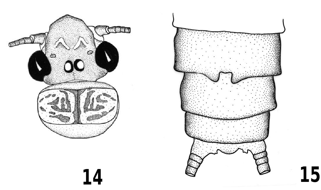
1 Tergum 8 with a small median lobe ( Fig. 15 View Figs ) ……………………………………………………… 2
1’ Tergum 8 unmodified ( Fig. 1 View Figs ) …..………………. 6
2 Aedeagal sac tubular, gradually tapered; basolateral area of sac with a pair of spiny patches ( Figs. 6 View Figs , 26 View Figs ) ………………………………………… 3
2’ Aedeagal sac with additional lobes; basolateral area of sac without spiny patches ( Figs. 2 View Figs , 17 View Figs ) ………………………………………………...…… 5
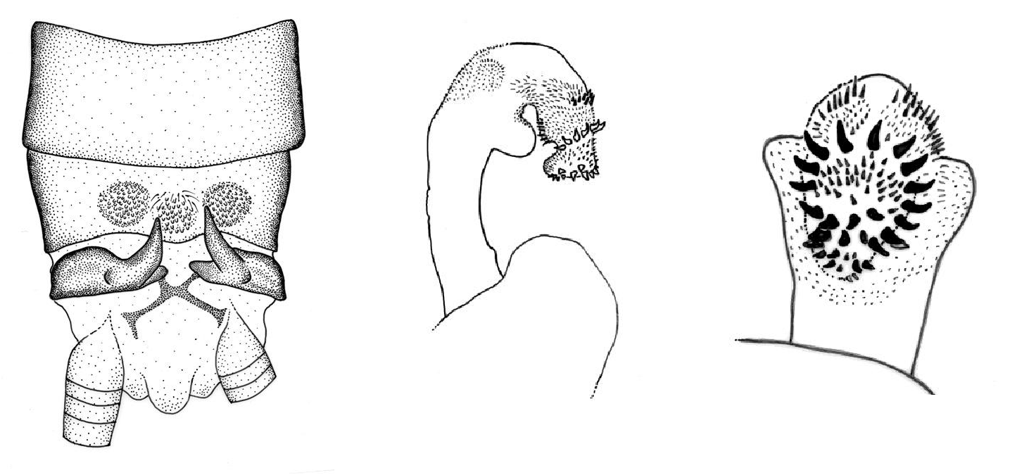
3 Largest spines on aedeagal sac form a transverse subapical row ( Fig. 26 View Figs ) ………………….... uchidai View in CoL
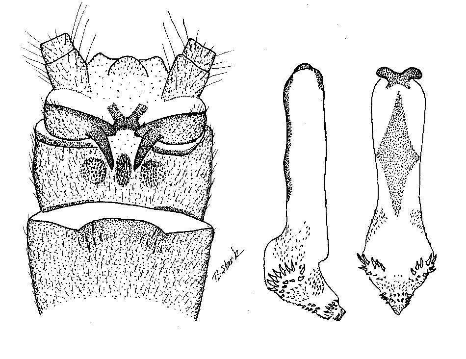
3’ Largest spines on aedeagal sac form irregular longitudinal rows ( Fig. 6 View Figs ) ………………………. 4
4 Largest spines on aedeagal sac located nearer base of sac; spines become smaller nearer apex ……………………………………………………. lisu View in CoL
4’ Largest spines clustered near tip of aedeagal sac ( Fig. 6 View Figs ) ……………………………………….. huang View in CoL
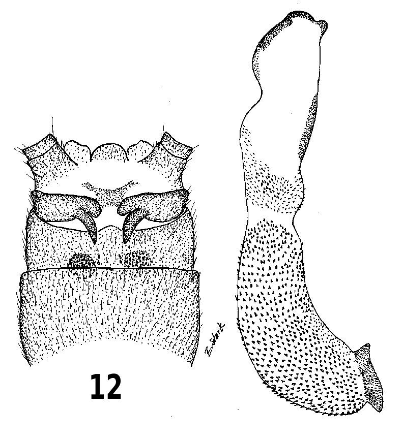
5 Largest aedeagal spines form transverse row more or less in center of apical spine patch ( Fig. 2 View Figs ) …………………………………………….. doisuthep View in CoL
5’ Largest aedeagal spines form transverse row at basal margin of apical spine patch ( Fig. 17 View Figs ) …………………………………………………. lobata View in CoL
6 Aedeagal armature entirely of small to moderate size spines ………………………….…………….. 7
6’ Aedeagal armature includes large black cultriform spines ……………………………….. 10
7 Everted aedeagal sac with one or more membranous lobes ………………………………. 8
7’ Everted aedeagal sac tubular, without extra lobes ……………………………………………………… 9
8 Mesal area of tergum 9 with two small irregular rows of sensilla between lateral patches; mesal section of aedeagal sac with a single finger-like lobe …………………………………………….... lao View in CoL
8’ Mesal area of tergum 9 without median sensilla between lateral patches; mesal section of aedeagal sac with a pair of finger-like lobes ……… imitatrix View in CoL
9 Major aedeagal armature on everted sac restricted to patch covering less than half sac length; basal half of sac constricted between sac apex and tube …………………………………………..….. simplex View in CoL
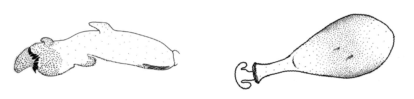
9’ Major aedeagal armature on everted sac covers almost entire sac length ( Fig. 13 View Figs ); base of sac constricted but sac gradually widens from base to subapical area ……………………….…….. hubleyi View in CoL
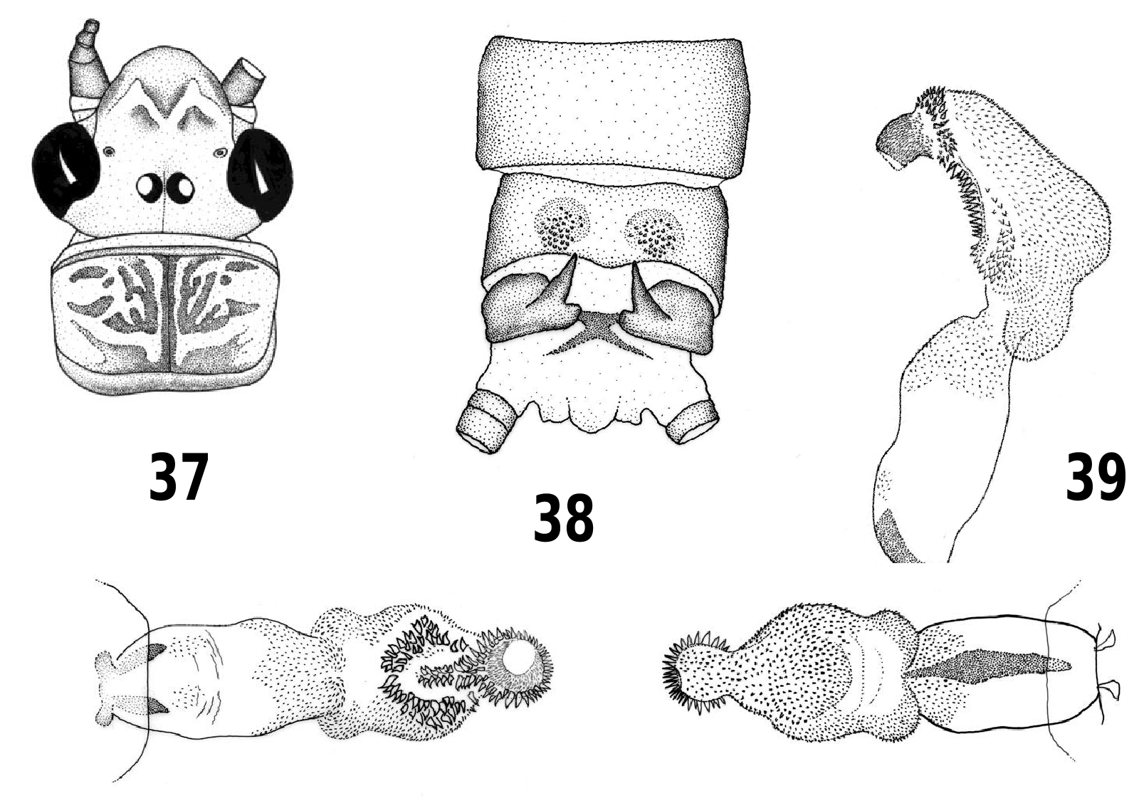
10 Subapical area of everted aedeagal sac bearing a complete single or double ring of large, black, cultriform spines (Figs. 21,39); tergum 9 with two large patches of sensilla, or a single fused median patch ( Fig. 20 View Figs ) …………………………………... 11
10’ Subapical area of everted aedeagal sac bearing an incomplete ring of large, black, cultriform spines; tergum 9 with median sensilla between lateral patches …………………………………………... 13
11 Almost entire lateral surface of aedeagal sac and apex of tube covered with a continuous spine patch ( Fig. 39 View Figs ); largest aedeagal spines form a complete subapical double ring; sensilla patches of tergum 9 widely separated ( Fig. 38 View Figs ) …… wieng View in CoL
11’ About half of aedeagal sac surface covered with continuous spine patch ( Fig. 19 View Figs ); largest aedeagal spines form a subapical single ring; sensilla patches of tergum 9 narrowly separated or united at base ( Fig. 20 View Figs ) ………………………………….. 12
12 Sensilla patches of tergum 9 completely divided; membranous lobe of aedeagal sac not covered with spines ……………………………… sertispina View in CoL
12’ Sensilla patches of tergum 9 united basally ( Fig. 20 View Figs ); membranous lobes of aedeagal sac covered with spines ( Fig. 21 View Figs ) …………………… occipitalis View in CoL
13 Everted aedeagal sac tubular, without membranous lobes; tergum 9 with a single median sensilla patch …………………… malayana View in CoL
13’ Everted aedeagal sac bulbous and bearing a small finger-like membranous lobe; tergum 9 with median sensilla grouped in two irregular rows ………………………………………… vietnamensis View in CoL
Figs. 1-3. Phanoperla doisuthep. 1. Male terminalia, dorsal. 2. Fully everted aedeagus, lateral. 3. Aedeagal sac apex.
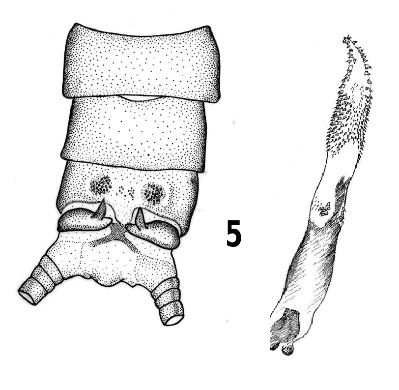
Figs. 4-7. Phanoperla huang. 4. Head and pronotum. 5. Male terminalia, dorsal. 6. Aedeagus, oblique ventrolateral. 7. Female terminalia.
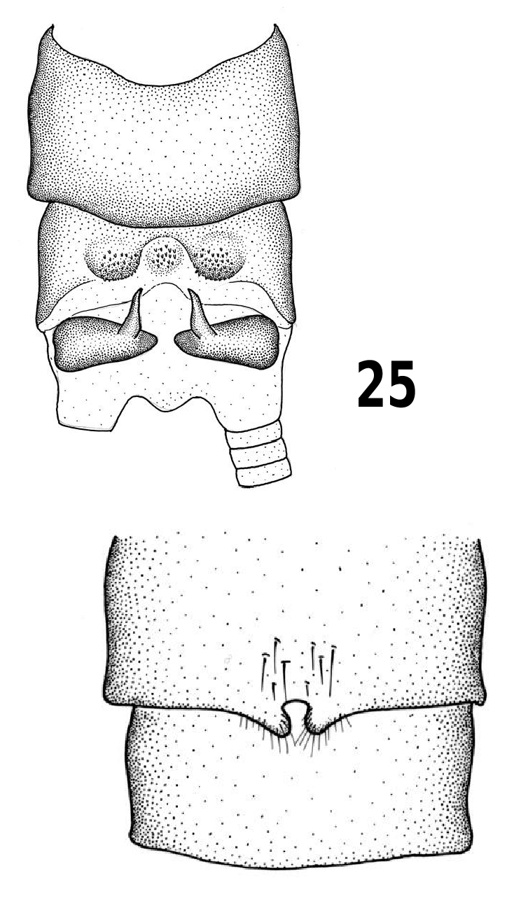
Figs. 24-29. Phanoperla uchidai. 24. Head and pronotum. 25. Male terminalia, dorsal. 26. Aedeagus, lateral. 27. Aedeagus, dorsal. 28. Aedeagus, ventral. 29. Female terminalia.
Figs. 16-18. Phanoperla lobata. 16. Male terminalia, dorsal. 17. Fully everted aedeagus, lateral. 18. Fully everted aedeagus, dorsal.
Figs. 19-23. Phanoperla occipitalis. 19. Head and pronotum. 20. Male terminalia, dorsal. 21. Aedeagus, lateral. 22. Female terminalia. 23. Egg.
No known copyright restrictions apply. See Agosti, D., Egloff, W., 2009. Taxonomic information exchange and copyright: the Plazi approach. BMC Research Notes 2009, 2:53 for further explanation.
|
Kingdom |
|
|
Phylum |
|
|
Class |
|
|
Order |
|
|
Family |