
Atractides Koch, 1836
|
publication ID |
https://doi.org/10.5281/zenodo.172007 |
|
publication LSID |
lsid:zoobank.org:pub:CDD71997-D63A-41B6-A3F8-5F50EBB416E8 |
|
persistent identifier |
https://treatment.plazi.org/id/03B187E0-FFD9-575B-CD09-F93D0C82CD19 |
|
treatment provided by |
Plazi (2016-04-04 09:30:58, last updated 2018-06-29 13:35:29) |
|
scientific name |
Atractides Koch, 1836 |
| status |
|
Genus Atractides Koch, 1836
Diagnosis: Character states as given for the family. Differential characters to separate Records ( Table 2): Benthos samples at nine spring sites in Gutland, 51 individuals ( Gerecke et al. 2005). E 1 129, E 7 54 larvae, parasitic on chironomids. 10 larvae were bred from a female from the spring site Lux Qu 19 (E 1). Attribution by rearing.
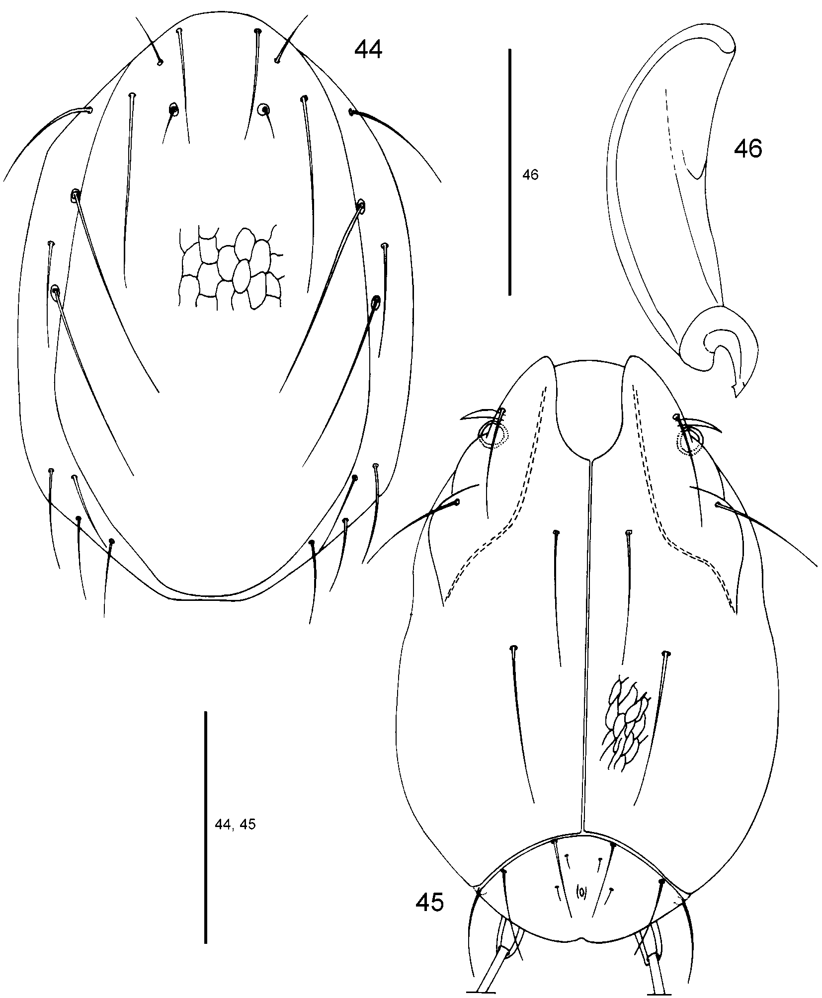
Description (unless otherwise indicated, n = 5): Idiosoma moderately elongated ( Figs. 44, 45 View FIGURES 44 – 46 ). Length/width of idiosoma 225–230 (227)/ 133–145 (140).
Dorsal idiosoma ( Fig. 44 View FIGURES 44 – 46 ): Dp relatively narrow and slightly tapered posteriorly, largest width in the posterior third of the plate, length/width 243–250 (247)/ 130–138 (134), Mp 2 Amdp 38–42 (40), Mp 1 Mp 1 46–50 (48), Mp 2 Mp 2 38–40 (39), Lp 1 Lp 1 34–36 (35), Lp 2 Lp 2 70–73 (72), Mp 1 Lp 1 12–14 (13), Mp 2 Lp 2 17–18 (17), Mp 1 Lp 2 20–23 (22), Lp 1 Lp 2 32–34 (33), Mp 1 18–22 (20), Mp 2 19–22 (21), Lp 1 47–65 (54), Lp 2 85–91 (89), Hu 57–67 (60), Mh 1 95–99 (97), Mh 2 88–96 (93), Mh 3 43–46 (44), Mh 4 37– 42 (40), Lh 1 43–52 (47), Lh 2 44–48 (46), Lh 3 45–48 (46).
Ventral idiosoma ( Fig. 45 View FIGURES 44 – 46 ): Expp relatively large (in contrast to the other Atractides species reported here). Length CXIIII (from the median gnathosomal bay to the posterior end of CXIIII) 180–190 (186), width CXIIII 75–79 (77), common median length of both CXIIII 160–165 (164), maximum length of CXIIII 225–230 (229), C 1 C 2 49–53 (51), C 1 Mmcp 15–17 (16), C 4 Pmcp 97–102 (100), C 1 C 4 53–59 (55), C 1 55–66 (60), C 2 47–52 (49), C 3 50–63 (56), C 4 95–100 (97), length/width Expp 44–46 (45)/ 79–87 (81), E 1 E 1 12 –16 (13), E 2 E 2 22 –23 (22), E 1 4 –6 (5), E 2 6 –8 (7), V 1 31–35 (32), V 2 34 – 37 (36), V 3 45 –51 (48), V 4 170–180 (176), V 1 V 1 23–24 (24), V 2 V 2 65 –70 (67), V 3 V 3 79 –82 (80), V 4 V 4 58 –66 (61), V 1 V 2 25–28 (26), length/width of projecting base of V 4 17–20 (18)/ 11 – 11 (11).
Gnathosoma: Base 72–76 (73), chelicera ( Fig. 46 View FIGURES 44 – 46 ) 67–72 (69), chela 18–19 (18), length/width P 1 + 2 31–34 (33)/ 29–33 (31), P 3 19–21 (20)/ 20–22 (21), length claw 11–13 (12), long seta on P 3 76–82 (79).
Legs: Leg I ( Fig. 47 View FIGURES 47 – 49 ): Total length 189–199 (194), length/height IL 1 (1 se) 30–32 (31)/ 18–20 (19), IL 2 (7 se) 37–39 (38)/ 17–18 (17), IL 3 (7 se) 32–35 (33)/ 16–17 (17), IL 4 (4 se, 1 so, 1 eu) 40–41 (40)/ 17–18 (17), IL 5 (12 se, 1 so, 2 eu) 50–52 (51)/ 14–15 (14).
Leg II ( Fig. 48 View FIGURES 47 – 49 ): Total length 205–214 (209), length/height IIL 1 (1 se) 31–32 (32)/ 19– 21 (20), IIL 2 (7 se) 38–39 (38)/ 18–19 (18), IIL 3 (4 se, 1 so) 33–35 (34)/ 16–18 (17), IIL 4 (9 se, 1 so) 43–45 (44)/ 16–18 (16), IIL 5 (12 se, 1 so) 60–63 (61)/ 12–14 (13).
Leg III ( Fig. 49 View FIGURES 47 – 49 ): Total length 241–252 (247), length/height IIIL 1 (1 se) 38–40 (39)/ 16–18 (17), IIIL 2 (6 se) 40–43 (42)/ 16–18 (17), IIIL 3 (4 se, 1 so) 41–42 (41)/ 15–16 (16), IIIL 4 (8 se, 1 so) 56–58 (57)/ 16–17 (17), IIIL 5 (10 se) 66–69 (68)/ 13–14 (14).
Diagnostic characters of larvae: To date, the larva of Atractides fonticolus has remained undescribed. It differs from the other Atractides larvae here with respect to leg length, length of Dp and width of Expp.
Gerecke, R., Stoch, F., Meisch, C. & Schrankel, I. (2005) Die Fauna der Quellen und des hyporheischen Interstitials in Luxemburg. Unter besonderer Berucksichtigung der Acari, Ostracoda und Copepoda. Ferrantia - Travaux scientifiques du Musee national d'histoire naturelle, Luxembourg, 41, 1 - 134.

No known copyright restrictions apply. See Agosti, D., Egloff, W., 2009. Taxonomic information exchange and copyright: the Plazi approach. BMC Research Notes 2009, 2:53 for further explanation.
|
Kingdom |
|
|
Phylum |
|
|
Class |
|
|
Order |
|
|
Family |
1 (by plazi, 2016-04-04 09:30:58)
2 (by ImsDioSync, 2016-11-27 03:28:29)
3 (by ImsDioSync, 2016-11-27 03:33:13)
4 (by ImsDioSync, 2018-06-29 13:35:29)
5 (by ExternalLinkService, 2019-09-26 22:29:22)
6 (by ExternalLinkService, 2021-11-09 17:19:20)
7 (by ExternalLinkService, 2021-11-10 06:04:56)
8 (by ExternalLinkService, 2021-11-12 09:02:13)
9 (by plazi, 2023-10-25 06:29:53)